 Back
Gas Exchange and Circulation in Animals
Study Guide - Smart Notes
 Tailored notes based on your materials, expanded with key definitions, examples, and context.
Gas Exchange and Circulation
Introduction to Gas Exchange
Gas exchange is a fundamental process in animals, involving the uptake of oxygen (O2) and the removal of carbon dioxide (CO2). These gases must be exchanged continuously to support cellular respiration and ATP production. The respiratory and circulatory systems work together to ensure efficient transport of gases, nutrients, and wastes throughout the body.
Oxygen is required for mitochondrial ATP production.
Carbon dioxide is a waste product that must be expelled to maintain cellular function.
Steps of Gas Exchange
Gas exchange in animals involves five major steps:
Ventilation: Movement of air or water through a specialized gas-exchange organ (e.g., lungs, gills).
Diffusion at the Respiratory Surface: O2 moves from the environment into the blood; CO2 moves from the blood into the environment.
Circulation: Transport of dissolved gases throughout the body via the circulatory system.
Diffusion at the Tissues: O2 moves from blood into tissues; CO2 moves from tissues into blood.
Cellular Respiration: Cells use O2 and produce CO2, creating gradients that drive diffusion.
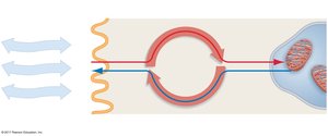
The Respiratory and Circulatory Systems
Organs of Gas Exchange
Animals have evolved various organs for gas exchange, depending on their size and environment:
Small animals may exchange gases directly across their moist body surfaces.
Larger or terrestrial animals require specialized organs (lungs, gills, tracheae) to increase surface area for diffusion.
Vertebrate Lungs
In vertebrates, lungs are the primary organs for gas exchange. Air enters through the mouth and nose, travels down the trachea, and branches into bronchi and bronchioles, ending in alveoli. Alveoli are tiny sacs that provide a large surface area and a thin barrier for efficient gas exchange.
Alveoli are lined with a thin aqueous film, epithelial cells, extracellular matrix, and capillary walls.
Humans have about 150 million alveoli per lung.
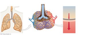
Mechanisms of Lung Ventilation
Lung ventilation varies among animal groups:
Positive pressure ventilation: Used by amphibians (e.g., frogs) to force air into the lungs.
Negative pressure ventilation: Used by mammals, including humans, where the diaphragm contracts to expand the chest cavity and draw air in.
During inhalation, the diaphragm moves down, lowering chest pressure and expanding the lungs. During exhalation, the diaphragm relaxes, reducing lung volume and expelling air.
Transport of Gases in Blood
Blood Composition and Function
Blood is a connective tissue composed of cells suspended in plasma. It transports gases, nutrients, wastes, hormones, and immune cells throughout the body.
Red blood cells (RBCs): Transport O2 and participate in CO2 transport.
White blood cells (WBCs): Part of the immune system.
Platelets: Involved in blood clotting.
Hemoglobin Structure and Function
Hemoglobin is the oxygen-carrying molecule in RBCs. Each hemoglobin molecule consists of four polypeptide chains, each with a heme group containing an iron ion (Fe2+) that binds O2. Each hemoglobin can carry up to four O2 molecules.
98.5% of O2 in blood is bound to hemoglobin; 1.5% is dissolved in plasma.
The rate of O2 unloading depends on the partial pressure of O2 in tissues.
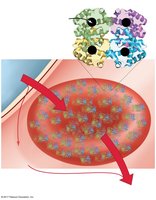
Circulatory Systems
Types of Circulatory Systems
Animals have evolved two main types of circulatory systems to transport gases and nutrients:
Open circulatory system: Hemolymph bathes organs directly; found in arthropods and most mollusks.
Closed circulatory system: Blood is contained within vessels; found in vertebrates, annelids, and cephalopods.
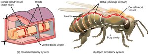
Blood Vessels
Blood vessels are classified based on their structure and function:
Arteries: Thick-walled vessels that carry blood away from the heart under high pressure.
Capillaries: Thin-walled vessels (one cell thick) where exchange of gases and nutrients occurs.
Veins: Thin-walled vessels that return blood to the heart; contain valves to prevent backflow.
The Lymphatic System
The lymphatic system collects excess interstitial fluid (lymph) and returns it to the circulatory system. It also absorbs fats from the digestive tract and plays a role in immune defense.
The Heart and Circulation
Heart Structure and Function
In closed circulatory systems, the heart is a muscular organ with chambers that pump blood through vessels. Most vertebrates have multi-chambered hearts, which allow separation of oxygenated and deoxygenated blood and support high metabolic rates.
Atrium (atria): Receives blood returning from circulation.
Ventricle: Pumps blood out to the body or lungs.
Valves ensure one-way flow and prevent backflow.
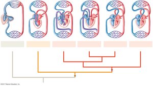
Evolution of Circulatory Circuits
Vertebrates have evolved increasingly complex hearts and circulatory circuits:
Fish: Two-chambered heart, single circuit.
Amphibians and most reptiles: Three-chambered heart, double circuit (pulmonary and systemic).
Birds and mammals: Four-chambered heart, complete separation of circuits.
Blood Flow in the Human Heart
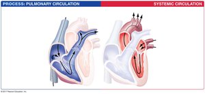
Blood flows through the heart in a specific sequence to maintain separation of oxygen-rich and oxygen-poor blood:
O2-poor blood enters the right atrium from the body (via venae cavae).
Moves to the right ventricle, then pumped to the lungs via the pulmonary artery.
O2-rich blood returns from the lungs to the left atrium (via pulmonary veins).
Moves to the left ventricle, then pumped to the body via the aorta.
The Cardiac Cycle and Blood Pressure
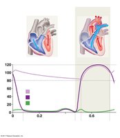
The cardiac cycle consists of systole (contraction) and diastole (relaxation) phases for both atria and ventricles. Blood pressure is measured as systolic (peak contraction) over diastolic (relaxation) pressure. Normal blood pressure is around 120/80 mm Hg; values above 140/90 mm Hg indicate hypertension.
High blood pressure increases risk of heart attack, stroke, and other cardiovascular diseases.
Regulation of Blood Pressure and Flow
Blood pressure and flow are regulated by baroreceptors in the heart and major arteries. When blood pressure drops, baroreceptors trigger responses to increase cardiac output, constrict arterioles, and shift blood volume to maintain flow to vital organs.
Respiratory and Cardiovascular Diseases
Respiratory Diseases
Chronic obstructive pulmonary disease (COPD): Long-term airflow obstruction.
Asthma: Allergen-induced constriction of bronchi.
Emphysema: Breakdown of alveolar walls, reduced elasticity.
Lung cancer: Rapidly metastasizing cancer, often linked to smoking.
Cardiovascular Diseases
Arteriosclerosis: Hardening and loss of elasticity in arteries.
Atherosclerosis: Accumulation of fatty plaques in arteries, leading to reduced blood flow and risk of clots (thrombus, embolus).
Myocardial infarction (heart attack): Blockage of coronary arteries, causing tissue death.
Cardiovascular disease is the leading cause of death worldwide; risk factors include age, tobacco use, poor diet, obesity, inactivity, and genetics.
Additional info: Effective prevention of cardiovascular disease involves lifestyle changes (diet, exercise, avoiding tobacco) and improved healthcare access.
