 Back
General Biology Lab Techniques: Safety, Measurement, Microscopy, and Spectrophotometry
Study Guide - Smart Notes
 Tailored notes based on your materials, expanded with key definitions, examples, and context.
Laboratory Safety and Preparation
Safety Rules and Laboratory Conduct
Laboratory safety is essential for preventing accidents and ensuring a productive learning environment. Students must adhere to strict safety protocols, including proper attire, chemical handling, and emergency procedures.
Dress Code: Closed-toe shoes, pants/skirts covering knees, and torso coverage are required. Eye protection must be worn when handling chemicals or glassware.
Chemical Handling: Read all labels carefully, use proper disposal methods, and never return chemicals to original containers.
Emergency Procedures: Know the location of eyewash stations, fire extinguishers, and report all accidents immediately.
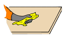
Cleanliness: Keep work areas clean and organized; clean glassware and surfaces before and after use.
Prohibited Activities: Eating, drinking, smoking, and bringing unauthorized persons into the lab are strictly forbidden.
Hazard Identification Systems
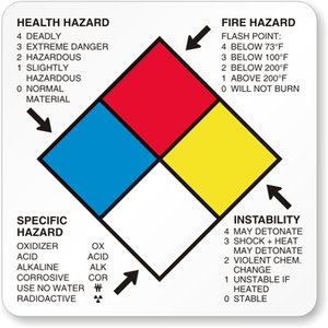
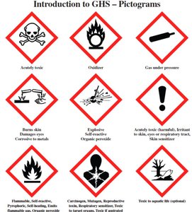
Understanding chemical hazards is critical. The NFPA label and Globally Harmonized System (GHS) pictograms provide information about health, fire, reactivity, and specific hazards.
NFPA Label: Uses a color-coded diamond to indicate hazard severity (0-4 scale) for health (blue), flammability (red), reactivity (yellow), and special hazards (white).
GHS Pictograms: Standardized symbols for toxicity, flammability, corrosivity, environmental hazards, and more.
Measurement Techniques and Metric System
Metric System Fundamentals
The metric system is the standard for scientific measurement, using units such as meter (m), liter (l), and gram (g). Prefixes indicate multiples or fractions of base units (e.g., kilo-, centi-, milli-, micro-, nano-).
Conversions: 1 meter = 100 centimeters = 1000 millimeters; 1 millimeter = 1000 micrometers; 1 micrometer = 1000 nanometers.
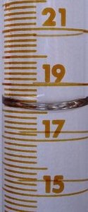
Accuracy: Always use the bottom of the meniscus when measuring liquids in graduated cylinders or pipettes.
Microscopy
Types of Microscopes
Microscopes are essential for studying cells and microorganisms. The three main types are:
Compound Light Microscope: Uses visible light and glass lenses; suitable for thin, transparent specimens; provides color images.
Electron Microscope: Uses electron beams and magnetic lenses; offers high resolution (up to 0.2 nm); images are black and white.
Stereomicroscope (Dissecting Scope): Provides three-dimensional views of opaque specimens using reflected light.
Parts and Operation of the Compound Microscope
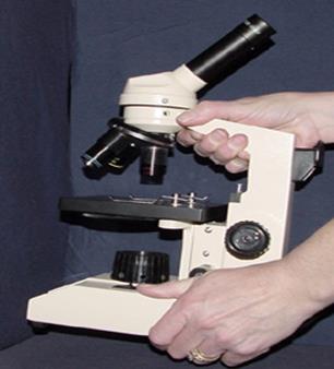
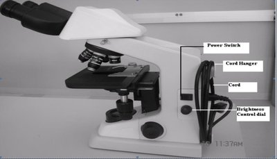
Proper handling and identification of microscope components are crucial for accurate observation.
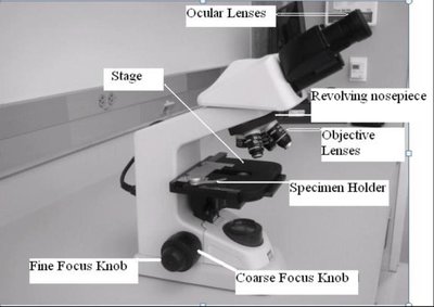
Ocular Lenses: Eyepieces, typically 10x magnification.
Objective Lenses: Multiple lenses (4x, 10x, 40x, 100x) for varying magnification.
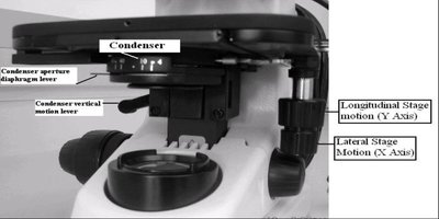
Stage: Platform for holding slides.
Coarse and Fine Focus Knobs: Adjust stage height and focus.
Condenser and Iris Diaphragm: Control light intensity and focus.
Microscope Focusing and Magnification
Focusing involves starting with the lowest magnification (4x scanning objective), centering the specimen, and gradually increasing magnification. The microscope is parfocal, meaning the image remains nearly focused when switching objectives.
Total Magnification Formula: Multiply ocular lens magnification by objective lens magnification.
Example:
Field of View (FOV) and Depth of Field
The field of view is the visible area through the microscope. As magnification increases, FOV decreases. Depth of field is the thickness of the specimen in focus; it also decreases with higher magnification.
FOV Calculation:
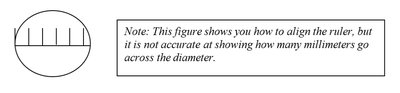
Example: If FOV at 4x is 4.5 mm, FOV at 10x is mm.
Estimating Cell Size
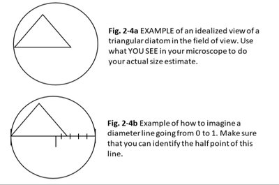
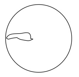
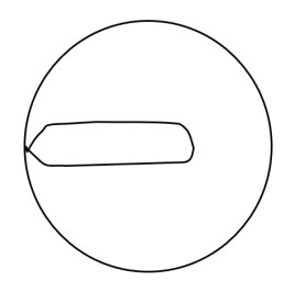
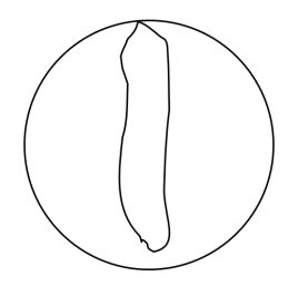
Cell size can be estimated by comparing the fraction of the FOV occupied by the cell and multiplying by the FOV diameter.
Example Calculation: If FOV at 40x is 450 µm and cell occupies 2/3, then µm.
Measurement Techniques & Spectrophotometry
Using Pipettes and Micropipettes
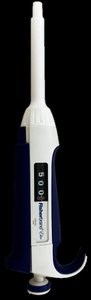
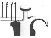
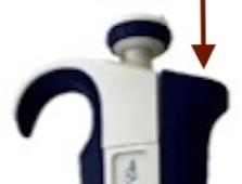
Accurate measurement of liquids is achieved using glass pipettes, micropipettes, and graduated cylinders. Micropipettes are used for volumes in microliters (µl).
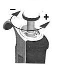
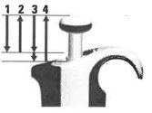
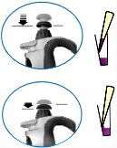
Micropipette Operation: Attach tip, set volume, aspirate sample to first stop, discharge to second stop, eject tip.
Volume Setting: Never exceed stated range; read display carefully.
Dilution Calculations
Dilutions are performed by mixing a specific volume of stock solution with solvent. The dilution fold and final concentration are calculated as follows:
Dilution Fold:
Final Concentration:
Preparation Formula:
Spectrophotometry and Beer’s Law
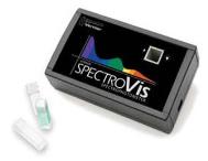
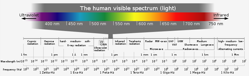
Spectrophotometry measures the absorbance of light by a solution to determine concentration. Beer’s Law states that absorbance is directly proportional to concentration.
Beer’s Law: Where A is absorbance, ε is molar absorptivity, l is path length, and c is concentration.
Calibration: Use a blank solution to set zero absorbance.
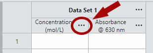
Data Collection: Measure absorbance at the wavelength of maximum absorption; plot absorbance vs. concentration.
Summary Table: Comparison of Microscopes
Microscope Type | Energy Source | Resolution | Color | Specimen Type |
|---|---|---|---|---|
Compound Light | Visible Light | ~0.1 µm | Color | Thin, transparent |
Electron | Electron Beam | ~0.2 nm | Black & White | Thin, fixed |
Stereomicroscope | Visible Light (Reflected) | Lower | Color | Opaque, 3D |
Key Terms and Concepts
Meniscus: The curve at the surface of a liquid in a measuring device; always read at the lowest point.
Magnification: The process of enlarging the appearance of an object.
Resolution: The ability to distinguish two points as separate.
Contrast: The degree to which specimen details stand out against the background.
Absorbance: The amount of light absorbed by a solution, measured by a spectrophotometer.
Beer’s Law: Relationship between absorbance and concentration.
Additional info: These notes expand on brief points in the lab manual to provide academic context, definitions, and formulas for self-contained study.
