 Back
BackGuided Study: Key Concepts in Animal Diversity and Evolution
Study Guide - Smart Notes
 Tailored notes based on your materials, expanded with key definitions, examples, and context.
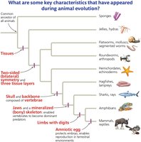
Q1. What are the key characteristics of animals described in Figure 27.1?
Background
Topic: Animal Evolution and Unique Traits
This question is testing your understanding of the evolutionary milestones and unique features that distinguish major animal groups. These characteristics are important for recognizing how animals have diversified and adapted over time.
Key Terms and Concepts:
Tissues: Groups of cells with a common structure and function.
Bilateral symmetry: Body plan with two mirror-image sides.
Three tissue layers: Ectoderm, mesoderm, endoderm.
Skull and backbone: Features of vertebrates.
Jaws and mineralized skeleton: Adaptations for feeding and support.
Limbs with digits: Key feature of tetrapods.
Amniotic egg: Adaptation for terrestrial life.
Step-by-Step Guidance
Examine the evolutionary tree in Figure 27.1 and note the order in which key characteristics appear as you move from sponges to mammals and reptiles.
Identify which groups first show the presence of tissues, and which groups develop bilateral symmetry and three tissue layers.
Observe where the skull and backbone (vertebrate traits) emerge, and which groups develop jaws and a mineralized skeleton.
Look for the appearance of limbs with digits and the amniotic egg, noting which animal groups possess these features.
Try solving on your own before revealing the answer!
Final Answer:
The key characteristics described in Figure 27.1 are: tissues, bilateral symmetry and three tissue layers, skull and backbone, jaws and mineralized skeleton, limbs with digits, and amniotic egg. These features mark major evolutionary milestones in animal diversity.
Each characteristic represents a significant adaptation that allowed animals to exploit new environments and ecological niches.
Q6. Label the figure of the sponge and describe the structure’s role. Draw the flow of water through the sponge.
Background
Topic: Sponge Anatomy and Water Flow
This question is testing your understanding of sponge structure and function, specifically how water moves through the sponge and the role of each anatomical feature.
Key Terms:
Choanocytes: Cells that create water currents and capture food.
Amoebocytes: Cells involved in digestion and distribution of nutrients.
Osculum: Large opening where water exits the sponge.
Pores: Small openings where water enters.
Step-by-Step Guidance
Identify and label the main structures in the sponge diagram: pores, choanocytes, amoebocytes, osculum, and canal system.
Describe the function of each labeled structure, focusing on how they contribute to water flow and feeding.
Trace the path of water: it enters through pores, moves through the canal system, passes by choanocytes, and exits via the osculum.
Explain how choanocytes generate water currents and capture food particles, and how amoebocytes distribute nutrients.
Try solving on your own before revealing the answer!
Final Answer:
The labeled sponge diagram should include pores (water entry), choanocytes (food capture and water movement), amoebocytes (nutrient distribution), and osculum (water exit). Water flows from the pores, through the canal system, past choanocytes, and out the osculum.
This flow is essential for feeding, respiration, and waste removal in sponges.
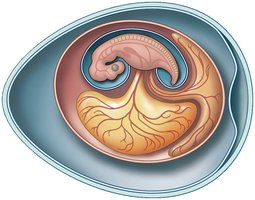
Q52. Label the four extraembryonic membranes in an amniotic egg and explain the role of each one.
Background
Topic: Amniotic Egg Structure and Function
This question is testing your knowledge of the adaptations that allow amniotes (reptiles, birds, mammals) to reproduce on land, focusing on the specialized membranes in the amniotic egg.
Key Terms:
Amnion: Membrane that surrounds and protects the embryo.
Chorion: Membrane involved in gas exchange.
Yolk sac: Provides nutrients to the embryo.
Allantois: Handles waste and gas exchange.
Step-by-Step Guidance
Examine the diagram of the amniotic egg and identify the four extraembryonic membranes: amnion, chorion, yolk sac, and allantois.
Label each membrane and describe its location relative to the embryo and the egg shell.
Explain the function of each membrane: protection, nutrition, waste management, and gas exchange.
Consider how these adaptations allow amniotes to reproduce away from water.
Try solving on your own before revealing the answer!
Final Answer:
The four extraembryonic membranes are: amnion (protects embryo), chorion (gas exchange), yolk sac (nutrients), and allantois (waste and gas exchange). Each membrane plays a crucial role in supporting embryonic development on land.
These adaptations are key to the success of amniotes in terrestrial environments.
