 Back
BackHeredity and Cellular Reproduction: Chromosomes, Mitosis, and Meiosis
Study Guide - Smart Notes
 Tailored notes based on your materials, expanded with key definitions, examples, and context.
Introduction to Heredity and Cellular Reproduction
Overview of Heredity
Heredity is the process by which genetic information is passed from one generation to the next. This process is fundamental to the continuity of life and is governed by the structure and function of DNA, genes, and chromosomes. Understanding heredity involves exploring how cells and organisms reproduce, how traits are inherited, and the molecular mechanisms underlying these processes.
DNA: The molecule that carries genetic information and is capable of being passed on to offspring.
Gene: A unit of heredity; a functional segment of DNA that can be transcribed.
Allele: A variant of a gene at a particular locus.
Chromosome: A structure made of DNA and proteins that carries genetic material.
Genome: The complete set of genetic information in an organism.
Genetics: The study of heredity and the form and function of genetic materials.
Chromosome Structure and Organization
Chromosome Composition
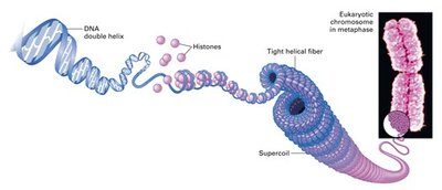
Chromosomes are cellular structures that carry genetic material. In eukaryotes, chromosomes are found in the nucleus and consist of linear double-stranded DNA molecules complexed with proteins called histones. In prokaryotes, chromosomes are typically circular and not enclosed by a nuclear membrane.
Histones: Proteins that help package DNA into a compact, organized structure.
Supercoiling: The further coiling of DNA to fit within the cell nucleus.
Key Chromosome Terms
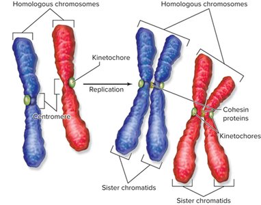
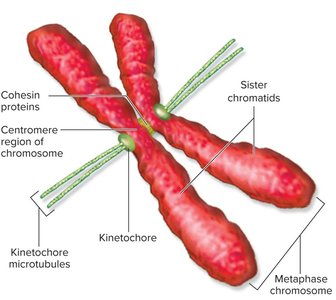
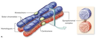
Chromatid: One of two identical halves of a replicated chromosome.
Sister chromatids: Two identical chromatids joined at the centromere, formed during DNA replication.
Non-sister chromatids: Chromatids of homologous chromosomes that are not identical.
Homologous chromosomes: Chromosome pairs, one from each parent, with matching loci.
Locus: A specific location on a chromosome, often corresponding to a gene.
Cell Division in Prokaryotes: Binary Fission
Process of Binary Fission
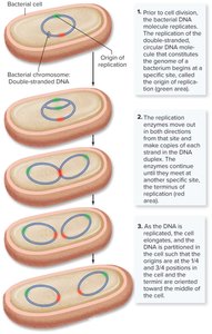
Binary fission is the method by which prokaryotic cells, such as bacteria, reproduce. It involves the replication of the circular DNA chromosome, segregation of the DNA, and division of the cell into two genetically identical daughter cells.
DNA replication begins at the origin of replication and proceeds bidirectionally.
The cell elongates, and the replicated DNA molecules move to opposite ends of the cell.
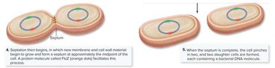
A septum forms, dividing the cell into two.
Eukaryotic Cell Cycle and Division
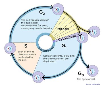
The Eukaryotic Cell Cycle
The cell cycle is the series of events that take place in a eukaryotic cell leading to its division and duplication. It consists of interphase (G1, S, G2 phases) and the mitotic phase (M phase), which includes mitosis and cytokinesis.
G1 phase: Primary growth phase; cell increases in size and prepares for DNA replication.
S phase: DNA synthesis; chromosomes are replicated.
G2 phase: Preparation for mitosis; organelles replicate, and microtubules organize.
M phase: Mitosis (nuclear division) and cytokinesis (cytoplasmic division).
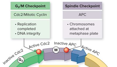
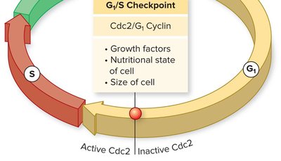
Cell Cycle Checkpoints
Checkpoints are control mechanisms that ensure the fidelity of cell division. They verify whether the processes at each phase of the cell cycle have been accurately completed before progression to the next phase. Failure of checkpoints can lead to uncontrolled cell division, resulting in tumors or cancer.
G1/S Checkpoint: Checks for cell size, nutrients, and DNA damage.
G2/M Checkpoint: Ensures DNA replication is complete and checks for DNA damage.
Spindle Checkpoint: Ensures all chromosomes are properly attached to the spindle before anaphase.
Mitosis: Nuclear Division in Eukaryotes
Purpose and Overview
Mitosis is the process by which a eukaryotic cell divides its nucleus, resulting in two genetically identical daughter cells. It is essential for growth, repair, and asexual reproduction in multicellular organisms.
Parent cell (2n) divides to form two daughter cells (2n), each genetically identical to the parent.
Stages: Prophase, Metaphase, Anaphase, Telophase.
Stages of Mitosis
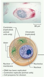
Interphase
During interphase, the cell prepares for division by replicating its DNA and organelles. Chromosomes are not yet visible as distinct structures.
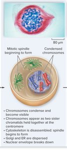
Prophase
Chromosomes condense and become visible. The nuclear envelope and nucleolus disassemble, and spindle fibers begin to form as centrosomes move to opposite poles.
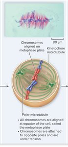
Metaphase
Chromosomes align at the metaphase plate, attached to spindle fibers from opposite poles via kinetochores. This alignment ensures equal segregation of chromosomes.
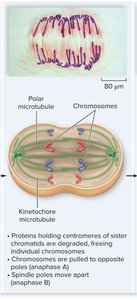
Anaphase
Centromeres split, and sister chromatids are pulled to opposite poles of the cell by shortening kinetochore microtubules. This ensures each daughter cell receives an identical set of chromosomes.
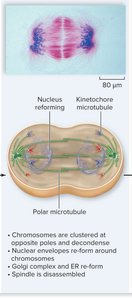
Telophase and Cytokinesis
Chromosomes decondense, nuclear envelopes reform around the two sets of chromosomes, and the spindle apparatus disassembles. Cytokinesis divides the cytoplasm, resulting in two separate daughter cells.
Karyotyping and Chromosome Analysis
Human Karyotype
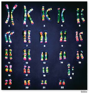
A karyotype is an organized profile of an individual's chromosomes, arranged by size and centromere position. Karyotyping is used for gene mapping and diagnosing chromosomal abnormalities.
Sexual Reproduction and Meiosis
Importance of Sexual Reproduction
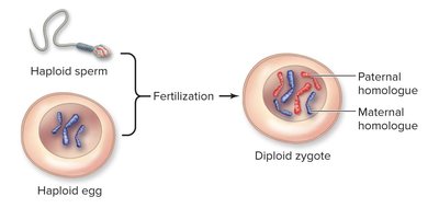
Sexual reproduction involves the formation of gametes (egg and sperm) through meiosis and their fusion during fertilization. This process increases genetic diversity by combining genetic material from two parents.
Diploid (2n): Cells with two sets of chromosomes (somatic cells).
Haploid (n): Cells with one set of chromosomes (gametes).
Meiosis: Reduction Division
Meiosis consists of two consecutive nuclear divisions (Meiosis I and II) that reduce the chromosome number by half, producing four genetically unique haploid cells from one diploid parent cell.
Meiosis I: Homologous chromosomes separate, crossing over occurs, and genetic variation is introduced.
Meiosis II: Sister chromatids separate, similar to mitosis.
Stages of Meiosis
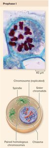
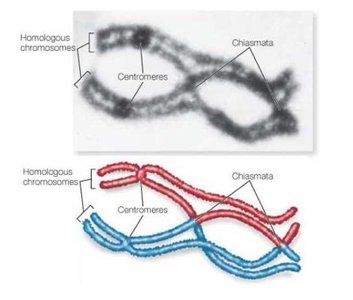
Prophase I
Chromosomes condense, homologous chromosomes pair up (synapsis), and crossing over occurs, resulting in genetic recombination between non-sister chromatids. Chiasmata are the sites of crossover.

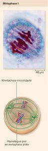
Metaphase I
Paired homologous chromosomes align at the metaphase plate. The orientation of each pair is random, contributing to genetic diversity through independent assortment.
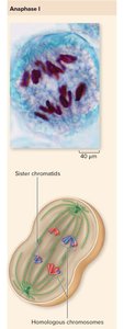
Anaphase I
Homologous chromosomes separate and move to opposite poles, while sister chromatids remain attached. This step reduces the chromosome number by half.
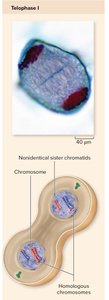
Telophase I and Cytokinesis
Nuclear envelopes may reform, and the cell divides. Each resulting cell is haploid, but chromosomes still consist of two chromatids.
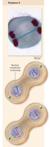
Meiosis II
Meiosis II resembles mitosis, where sister chromatids are separated, resulting in four haploid cells, each genetically distinct due to crossing over and independent assortment.
Comparison of Mitosis and Meiosis
Key Differences
Feature | Mitosis | Meiosis |
|---|---|---|
Number of divisions | 1 | 2 |
Number of daughter cells | 2 | 4 |
Chromosome number in daughter cells | Diploid (2n) | Haploid (n) |
Genetic identity | Identical to parent | Genetically unique |
Role | Growth, repair, asexual reproduction | Gamete production, genetic diversity |
Summary Table: Chromosome and Chromatid Numbers During Division
Stage | # Chromatids | # Chromosomes |
|---|---|---|
G1 | n | n |
G2 | 2n | n |
Metaphase | 2n | n |
Anaphase | n | n |
Metaphase I (Meiosis) | 2n | n |
Anaphase I (Meiosis) | n | n |
Metaphase II (Meiosis) | n | n |
Gametes | n | n |
Conclusion
Understanding heredity and cellular reproduction is essential for comprehending how genetic information is transmitted and how genetic diversity arises. The processes of mitosis and meiosis ensure the continuity of life and the variation necessary for evolution and adaptation.
