 Back
BackHuman Skeletal System: Structure, Function, and Evolutionary Context
Study Guide - Smart Notes
 Tailored notes based on your materials, expanded with key definitions, examples, and context.
Introduction to the Human Skeletal System
The human skeletal system is a complex framework of bones that provides structural support, protection for vital organs, and facilitates movement. In addition to these mechanical functions, bones play critical roles in blood cell production and mineral storage. Understanding the anatomy and function of individual bones is essential for studies in biology, anatomy, and evolutionary science.
Bone Structure and Composition
General Properties of Bone
Support and Movement: Bones provide a rigid framework that supports the body and acts as levers for muscle action.
Protection: Bones protect vital organs such as the brain (skull) and spinal cord (vertebral column).
Blood Cell Generation: Red marrow within bones is the site of hematopoiesis (blood cell formation).
Composition: Bone is a living tissue composed of a matrix of collagen fibers reinforced with minerals such as calcium, phosphate, and carbonate, giving it both strength and flexibility.
Analogy: Bone structure is often compared to steel-reinforced concrete due to its combination of toughness and rigidity.
Major Bones of the Human Skeleton
Axial and Appendicular Skeleton
Axial Skeleton: Includes the skull, vertebral column, and rib cage.
Appendicular Skeleton: Comprises the limbs and girdles (pectoral and pelvic) that attach them to the axial skeleton.
Pectoral Girdle and Upper Limb
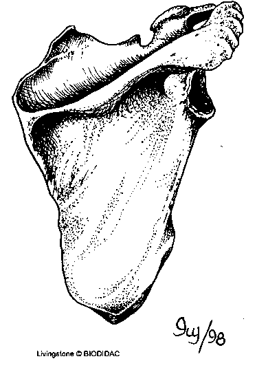
Scapula (Shoulder Blade): A triangular bone with a prominent ridge, palpable on the back. It serves as an attachment site for muscles and articulates with the clavicle and humerus.
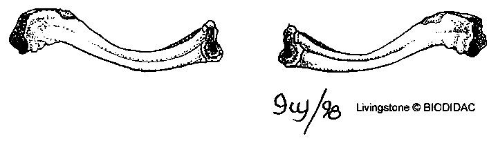
Clavicle (Collar Bone): An S-shaped bone with one rounded and one flat end, connecting the scapula to the sternum.
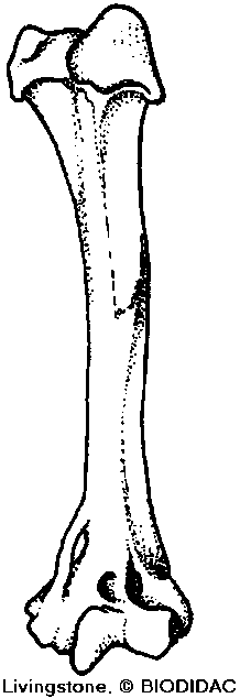
Humerus: The upper arm bone, with a ball-shaped head that articulates with the scapula and a spool-shaped end for the elbow joint. The humerus features a depression for the ulna during arm extension.
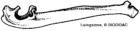
Ulna: One of the two forearm bones, characterized by a large, semicircular notch for articulation with the humerus.
Radius: The other forearm bone, with a round disk at one end allowing rotation of the forearm (pronation and supination).
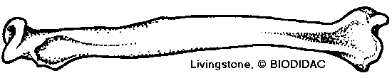
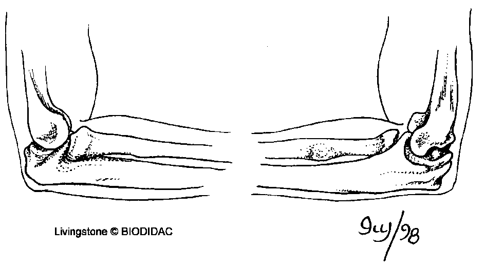
Elbow Joint: The humerus, ulna, and radius articulate to form the elbow, allowing flexion, extension, and rotation of the forearm.
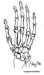
Carpals: Eight small bones forming the wrist, arranged in two rows.
Metacarpals: Five bones forming the palm of the hand.
Phalanges: Fourteen bones in the fingers (three per finger, two in the thumb).
Pelvic Girdle and Lower Limb
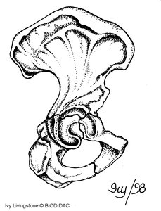
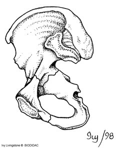
Os Coxa (Hip Bone): Formed by the fusion of the ilium, ischium, and pubis. The large socket (acetabulum) articulates with the femur. The structure reflects evolutionary changes from lower vertebrates.
Femur (Thigh Bone): The longest bone in the body, with a ball-and-neck structure fitting into the hip socket. Fractures of the neck are common in hip injuries.
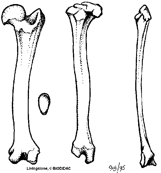
Patella (Knee Cap): A triangular bone that protects the knee joint and improves leverage for thigh muscles.
Tibia (Shin Bone): The larger, weight-bearing bone of the lower leg, with a sharp anterior ridge.
Fibula: The slender bone of the lower leg, providing muscle attachment and lateral stability. Contains trabeculae for strength with minimal weight.
Tarsals: Seven ankle bones, including the calcaneus (heel bone) and astragalus (talus), which articulate with the tibia.
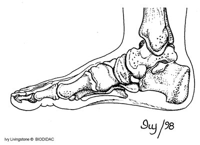
Metatarsals: Five long bones of the foot.
Phalanges: Toe bones, with the same naming as finger bones.
Skull and Associated Bones
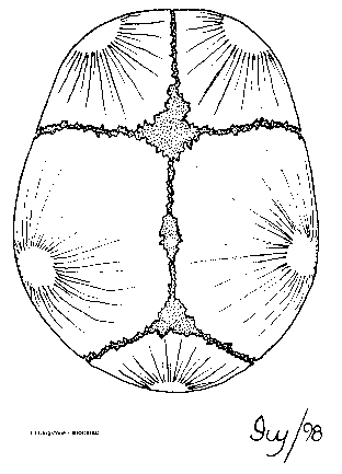
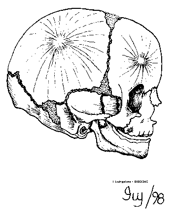
Skull: Composed of 29 bones, including those of the cranium, face, middle ear, and hyoid. At birth, the bones are not fully fused, leaving soft spots called fontanels.
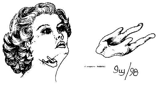
Hyoid: A horseshoe-shaped bone under the chin, supporting the tongue and associated muscles.
Vertebral Column
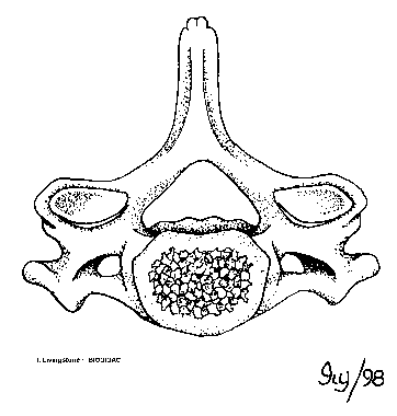
Cervical Vertebrae: Seven neck vertebrae, each with a spool-shaped body and foramina for blood vessels. The atlas (C1) supports the skull and allows nodding; the axis (C2) has a tooth-like projection (dens) for head rotation.
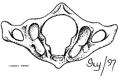
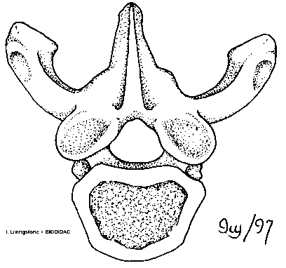
Thoracic Vertebrae: Twelve vertebrae attached to ribs, identified by rib facets.
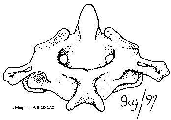
Lumbar Vertebrae: Five lower back vertebrae, largest in size, lacking rib facets and foramina.
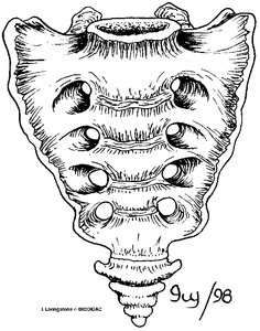
Sacrum: Five fused vertebrae forming a single bone, articulating with the os coxae.
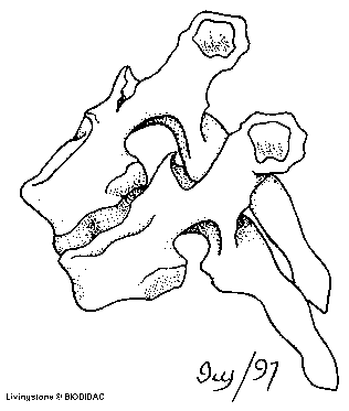
Coccyx: Vestigial tail vertebrae, usually fused into one or two bones.
Thoracic Cage
Sternum: The breastbone, consisting of the manubrium, body, and xiphoid process.
Ribs: Twelve pairs; the first seven are "true" ribs (directly attached to the sternum), the next five are "false" ribs (indirectly attached or floating).
Costal Cartilages: Connect ribs to the sternum, visible only in articulated skeletons.
Intervertebral Discs: Fibrocartilaginous pads between vertebrae, providing cushioning and flexibility. Herniation can compress spinal nerves.
Comparative Anatomy and Evolutionary Context
Pelvic Girdle Sexual Dimorphism
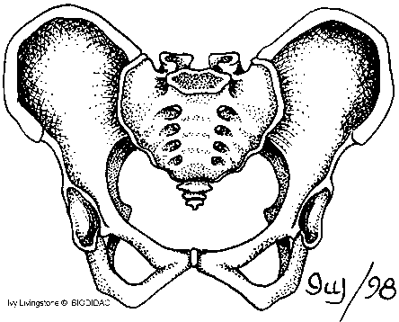
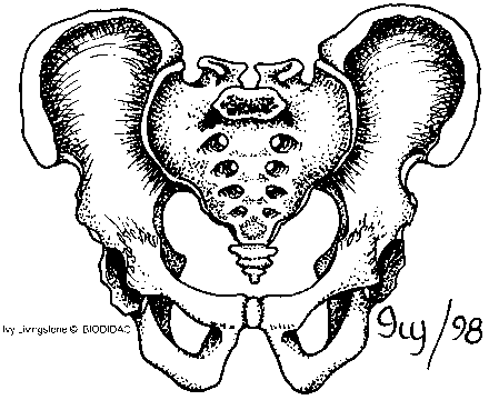
Male vs. Female Pelvis: The female pelvis has a larger, rounder pelvic inlet and a wider pubic arch angle, adaptations for childbirth. The male pelvis is more heart-shaped with a narrower pubic arch.
Evolutionary Insights from Bones
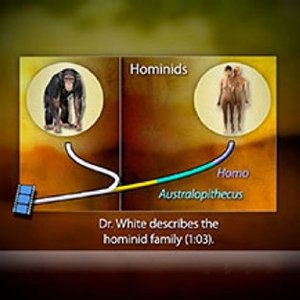
Phylogenetic Trees: Used to illustrate evolutionary relationships among species.
Hominids (Hominins): The group that includes modern humans and their extinct relatives, such as Australopithecus.
Comparative Anatomy: Features such as the pelvis, teeth, and feet provide evidence for evolutionary relationships between humans, chimpanzees, and extinct hominids.
Key Terms and Definitions
Foramen: A hole in a bone, typically for the passage of nerves or blood vessels.
Trabeculae: Sponge-like network of bone tissue providing strength with minimal weight.
Fontanel: Soft spot on a baby's skull where bones have not yet fused.
Articulation: The joint or connection between bones.
Summary Table: Major Human Bones and Their Features
Bone | Location | Key Features |
|---|---|---|
Scapula | Shoulder | Triangular, palpable ridge |
Clavicle | Shoulder | S-shaped, connects scapula to sternum |
Humerus | Upper arm | Ball-and-socket joint, depression for ulna |
Ulna | Forearm (medial) | Semicircular notch, forms elbow tip |
Radius | Forearm (lateral) | Round disk for rotation |
Femur | Thigh | Largest bone, ball-and-neck structure |
Tibia | Lower leg (medial) | Sharp anterior ridge |
Fibula | Lower leg (lateral) | Slender, supports muscles |
Patella | Knee | Triangular, protects knee joint |
Os Coxa | Pelvis | Fusion of ilium, ischium, pubis |
Sacrum | Pelvis | Five fused vertebrae |
Coccyx | Pelvis | Vestigial tailbone |
Skull | Head | 29 bones, fontanels in infants |
Vertebrae | Spine | Cervical (7), thoracic (12), lumbar (5) |
Ribs | Thorax | 12 pairs, true/false/floating |
Sternum | Thorax | Manubrium, body, xiphoid process |
Additional info:
Bone identification is essential for understanding human anatomy, evolutionary biology, and comparative anatomy with other vertebrates.
Sexual dimorphism in the pelvis is a key adaptation for childbirth in humans.
Comparative studies of bones (e.g., pelvis, teeth, feet) provide evidence for evolutionary relationships among hominids.
