 Back
BackCh. 7 part 2 (7.4-7.6)
Study Guide - Smart Notes
 Tailored notes based on your materials, expanded with key definitions, examples, and context.
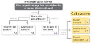
Cell Systems Overview
Introduction to Cell Systems
Cellular life is defined by the collaboration of internal structures, each contributing to the cell's overall function. The main systems discussed here are nuclear transport, the endomembrane system, and the dynamic cytoskeleton, which together orchestrate the movement, processing, and organization of molecules within eukaryotic cells.
Nuclear Transport (7.4)
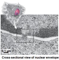
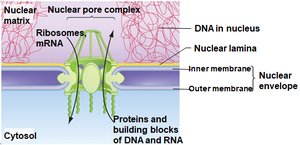
Structure and Function of the Nuclear Envelope
The nuclear envelope is a double-membrane structure that surrounds the nucleus, separating it from the cytosol. It is perforated with nuclear pores, which regulate the movement of molecules between the nucleus and cytoplasm.
Double Membrane: Consists of an inner and outer membrane.
Nuclear Pores: Complexes that allow selective transport of molecules.
Nuclear Lamina: Provides structural support to the envelope.
Mechanisms of Nuclear Transport
Transport through nuclear pores is highly selective and energy-dependent. Small molecules like nucleotides can diffuse freely, but larger molecules such as proteins, ribosomes, and RNA require specific signals.
Nuclear Localization Signal (NLS): Proteins destined for the nucleus must have an NLS, a molecular address tag.
Nuclear Export Signal (NES): Ribosomes and mRNA exiting the nucleus require an NES.
Selective Transport: Only molecules with the correct tags are actively transported.

Endomembrane System (7.5)
Overview and Secretory Pathway
The endomembrane system is responsible for manufacturing, shipping, and recycling cellular cargo. It includes the endoplasmic reticulum (ER), Golgi apparatus, vesicles, and lysosomes. The secretory pathway is a model describing the stepwise movement of proteins from synthesis to secretion.
Rough ER: Site of protein synthesis and initial processing.
Golgi Apparatus: Further modifies, sorts, and packages proteins.
Vesicles: Transport proteins between organelles and to the plasma membrane.
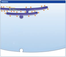


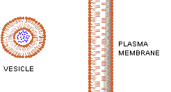
Vesicle Formation and Fusion
Membranes are fluid, allowing vesicles to form by pinching off and fuse with target membranes to deliver their contents. Vesicles move materials into, out of, and within the cell.
Endocytosis: Uptake of materials from outside the cell.
Exocytosis: Secretion of materials to the outside.
Intracellular Transport: Movement between organelles.
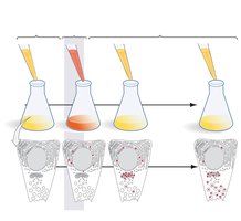
Pulse-Chase Experiment
The pulse-chase experiment is used to track the movement of proteins through the endomembrane system. Cells are exposed to radiolabeled amino acids (pulse), followed by unlabeled amino acids (chase), allowing visualization of protein trafficking.
Pulse: Short exposure to labeled amino acids.
Chase: Replacement with unlabeled amino acids.
Tracking: Proteins are followed as they move from ER to Golgi to vesicles.

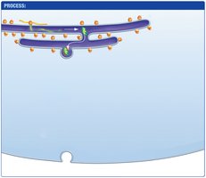
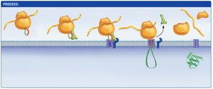
Protein Entry into the Endomembrane System: The Signal Hypothesis
Proteins destined for the endomembrane system begin synthesis on free ribosomes. The first amino acids form an ER signal sequence, which is recognized by the signal recognition particle (SRP). The SRP binds to its receptor on the ER membrane, and the protein is translocated into the ER lumen.
ER signal sequence is synthesized by ribosome.
SRP binds to signal sequence and halts synthesis.
SRP binds to SRP receptor on ER membrane.
SRP is released; protein enters ER via translocon.
Signal sequence is removed; protein synthesis completes.


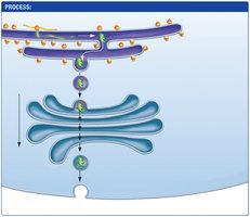
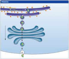
Protein Processing in the Golgi Apparatus
Proteins are further modified in the Golgi apparatus, where each cisterna contains different enzymes. Modifications occur as proteins move from the cis to the trans face, after which they are sorted and packaged for their final destination.
Stepwise Processing: Sequential modification in Golgi cisternae.
Protein Sorting: Proteins are tagged for specific destinations.
Vesicle Transport: Vesicles bud off and deliver proteins to their targets.


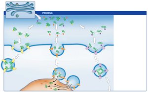
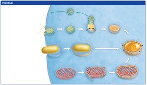
Lysosome Formation and Recycling
Lysosomes are recycling centers formed by three mechanisms: phagocytosis, autophagy, and receptor-mediated endocytosis. They degrade and recycle cellular components.
Phagocytosis: Uptake of large particles into phagosomes.
Autophagy: Degradation of damaged organelles via autophagosomes.
Receptor-mediated Endocytosis: Uptake of specific molecules via vesicles.
Dynamic Cytoskeleton (7.6)
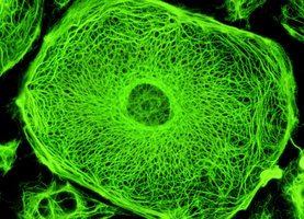
Structure and Types of Cytoskeletal Filaments
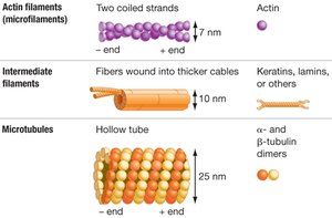
The cytoskeleton is a dynamic network of protein filaments that controls the location of organelles and is essential for cell shape, movement, and division. There are three main types of filaments: actin filaments, intermediate filaments, and microtubules.
Actin Filaments: Composed of actin; involved in cell movement and shape.
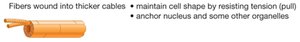
Intermediate Filaments: Composed of keratins, lamins, etc.; provide structural support.
Microtubules: Composed of tubulin dimers; involved in chromosome movement and intracellular transport.
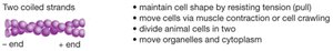
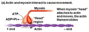
Actin Filaments and Cell Movement
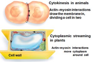
Actin filaments interact with myosin to produce cell movements, including muscle contraction, cell crawling, cytokinesis, and cytoplasmic streaming.
Actin-Myosin Interaction: Myosin heads bind to actin, hydrolyze ATP, and cause filament sliding.
Cytokinesis: Actin-myosin interactions divide animal cells.
Cytoplasmic Streaming: Moves cytoplasm in plant cells.

Intermediate Filaments
Intermediate filaments anchor the nucleus and other organelles, maintaining cell shape by resisting tension.
Structural Support: Provide mechanical strength.
Anchoring: Secure organelles in place.
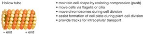
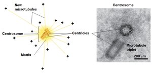
Microtubules
Microtubules are hollow tubes that move chromosomes during cell division, assist in cell plate formation, and provide tracks for intracellular transport. They are formed from α- and β-tubulin dimers.
Chromosome Movement: Essential during mitosis and meiosis.
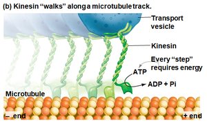
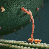
Intracellular Transport: Kinesin and other motor proteins move vesicles along microtubule tracks.
Cell Shape: Resist compression forces.
Kinesin and Intracellular Transport
Kinesin is a motor protein that "walks" along microtubule tracks, transporting vesicles and organelles. Each step requires ATP hydrolysis.
ATP Hydrolysis: Provides energy for movement.
Directional Transport: Moves cargo toward the plus end of microtubules.
Summary Table: Cytoskeletal Filaments
Filament Type | Structure | Main Functions |
|---|---|---|
Actin Filaments | Two coiled strands (7 nm) | Cell shape, movement, cytokinesis, organelle transport |
Intermediate Filaments | Fibers wound into cables (10 nm) | Structural support, anchoring organelles |
Microtubules | Hollow tubes (25 nm) | Chromosome movement, intracellular transport, cell shape |
Additional info: The notes expand on brief points to provide academic context, including definitions, examples, and stepwise explanations of cellular processes.
