 Back
Inside the Cell: Structure and Function of Prokaryotic and Eukaryotic Cells
Study Guide - Smart Notes
 Tailored notes based on your materials, expanded with key definitions, examples, and context.
Inside the Cell: Structure and Function
Introduction
This study guide covers the fundamental structures and functions of prokaryotic and eukaryotic cells, as outlined in Chapter 7.1–7.3. Understanding cell structure is essential for grasping how life’s properties emerge from the collaboration of internal cellular components.

Cell Theory and Classification
Cell Theory
Cell theory states that all organisms are composed of cells, which are the basic units of life. All cells share several key features:
Proteins: Perform most cellular functions.
Nucleic acids: Store, transmit, and process genetic information.
Carbohydrates: Provide energy, carbon, support, and identity.
Plasma membrane: Serves as a selectively permeable barrier.
Types of Cells
Prokaryotes: Lack a membrane-bound nucleus (Domains: Bacteria and Archaea).
Eukaryotes: Have a membrane-bound nucleus (Domain: Eukarya).
Prokaryotic Cell Structure
Overview of Prokaryotic Cells
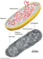
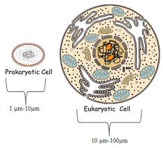
Prokaryotic cells, including bacteria and archaea, are structurally diverse and more complex than previously thought. They are typically 1–10 µm in diameter.
Key Components of Prokaryotic Cells
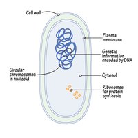
Chromosome: Usually a single, circular DNA molecule located in the nucleoid region, associated with proteins for structural support.
Plasmids: Small, circular DNA molecules that replicate independently and often carry advantageous genes.
Ribosomes: Macromolecular machines for protein synthesis, composed of RNA and protein subunits.
Cytoplasm: All contents within the plasma membrane.
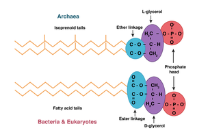
Plasma membrane: Phospholipid bilayer with proteins; composition differs between bacteria and archaea.
Cell wall: Provides structural support; in bacteria, primarily composed of peptidoglycan.
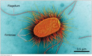
External structures: Flagella (motility) and fimbriae (attachment).
Organization of Genetic Material
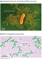
The chromosome is the most prominent structure in prokaryotic cells. DNA is often supercoiled to fit within the cell, and plasmids provide additional genetic capabilities.
Ribosomes in Prokaryotes
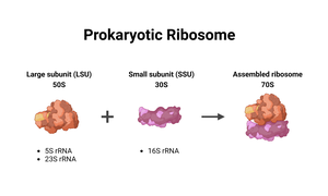
Prokaryotic ribosomes consist of a large (50S) and small (30S) subunit, forming a 70S ribosome. They are responsible for translating mRNA into proteins.
Cytoskeleton in Prokaryotes
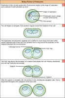
Long, thin protein filaments form a cytoskeleton, essential for cell division and maintaining cell shape.
Internal Membrane Complexes
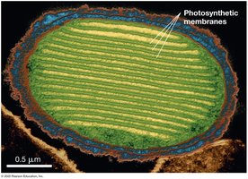
Some prokaryotes, especially photosynthetic species, have internal membrane systems that increase surface area for energy conversion.
Specialized Organelles in Prokaryotes
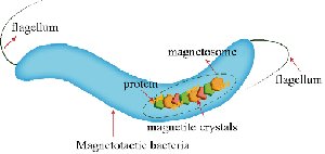
Certain bacteria possess organelles for specialized functions, such as storing ions or orienting with magnetic fields.
Cell Wall and External Structures
The cell wall acts as a protective exoskeleton. Many prokaryotes also have flagella for movement and fimbriae for attachment.
Eukaryotic Cell Structure
Overview of Eukaryotic Cells
Eukaryotic cells are generally larger (5–100 µm) and more complex than prokaryotic cells. They include protists, fungi, plants, and animals, and may be unicellular or multicellular.
Benefits of Compartmentalization
Separation of incompatible chemical reactions.
Increased efficiency of cellular processes.
The Nucleus
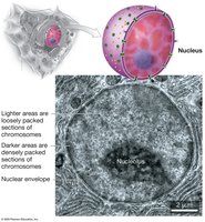
The nucleus is a large, membrane-bound compartment containing the cell’s genetic material. It is surrounded by a double membrane (nuclear envelope) and contains the nucleolus, where ribosomal RNA is synthesized.
Ribosomes in Eukaryotes
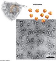
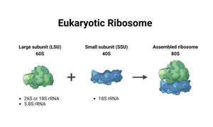
Eukaryotic ribosomes are larger (80S) than prokaryotic ribosomes and can be free in the cytosol or bound to the endoplasmic reticulum (ER).
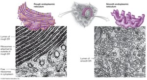
Endoplasmic Reticulum (ER)
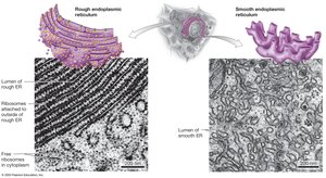
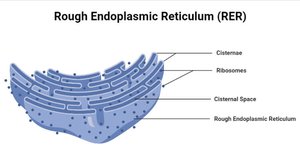
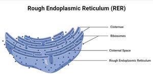
Rough ER (RER): Studded with ribosomes; synthesizes proteins for secretion or membrane insertion.
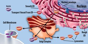
Smooth ER (SER): Lacks ribosomes; synthesizes lipids, detoxifies chemicals, and stores Ca2+ ions.
Golgi Apparatus
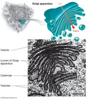
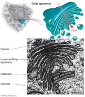
The Golgi apparatus processes, sorts, and ships proteins from the ER. It consists of stacked, flat membranous sacs (cisternae) with a cis (receiving) and trans (shipping) face.
Lysosomes
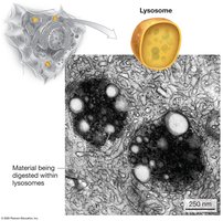
Lysosomes are recycling centers found in animal cells, containing acid hydrolases for breaking down macromolecules and cellular debris. They maintain an acidic pH for optimal enzyme activity.
Vacuoles
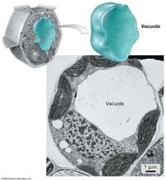
Vacuoles are prominent in plant and fungal cells, serving roles in storage, digestion, and protection. They may contain water, ions, proteins, pigments, or defensive compounds.
Peroxisomes
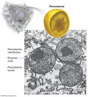
Peroxisomes are organelles that carry out oxidation reactions, breaking down fatty acids and detoxifying hydrogen peroxide via the enzyme catalase. Specialized peroxisomes in plants (glyoxysomes) convert fats to sugars.
Mitochondria
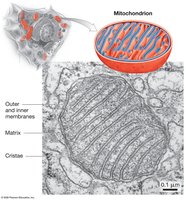
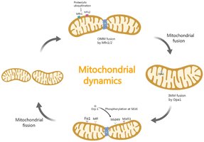
Mitochondria are the powerhouses of the cell, generating ATP through cellular respiration. They have a double membrane, with the inner membrane folded into cristae, and contain their own DNA and ribosomes.
Chloroplasts
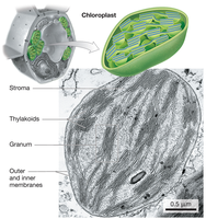
Chloroplasts are found in plants and algae, where photosynthesis occurs. They have three membranes, with thylakoids arranged in stacks (grana) and a surrounding stroma. Chloroplasts also contain their own DNA and ribosomes.
Cytoskeleton
The cytoskeleton is an extensive network of protein fibers that provides structural support, facilitates intracellular transport, and organizes cellular components.
Cell Wall and Extracellular Matrix
Fungi, algae, and plants have a rigid cell wall outside the plasma membrane for structural support. Animal cells lack a cell wall but are supported by an extracellular matrix (ECM) composed of proteins and polysaccharides.
Summary Table: Eukaryotic Cell Components
Organelle | Main Function | Present In |
|---|---|---|
Nucleus | Information storage and processing | All eukaryotes |
Ribosomes | Protein synthesis | All eukaryotes |
Rough ER | Protein synthesis and processing | All eukaryotes |
Smooth ER | Lipid synthesis and detoxification | All eukaryotes |
Golgi apparatus | Protein, lipid, and carbohydrate processing | All eukaryotes |
Lysosomes | Digestion and recycling | Animals |
Vacuoles | Storage, digestion, recycling | Plants, fungi |
Peroxisomes | Oxidation of fatty acids, detoxification | All eukaryotes |
Mitochondria | ATP production | All eukaryotes |
Chloroplasts | Photosynthesis | Plants, algae |
Cytoskeleton | Structural support, movement | All eukaryotes |
Cell wall | Protection, structural support | Plants, fungi, algae |
Integration of Cell Structure and Function
Structure-Function Relationship
The structure of each cell component is closely related to its function. For example, fat cells are specialized for lipid storage, while muscle cells are elongated for contraction. The number and type of organelles vary according to the cell’s role.
Dynamic Nature of Cells
Cells are dynamic, with organelles and molecules constantly moving and being replaced. Techniques such as differential centrifugation and fluorescence tagging allow researchers to study cell components in detail.
Additional info: The endosymbiosis theory explains the origin of mitochondria and chloroplasts as formerly free-living bacteria that established a symbiotic relationship with ancestral eukaryotic cells.
