 Back
BackIntroduction to Animal Diversity: Key Innovations, Porifera, and Cnidarians
Study Guide - Smart Notes
 Tailored notes based on your materials, expanded with key definitions, examples, and context.
Animal Diversity and Evolution
Overview of Animal Kingdom
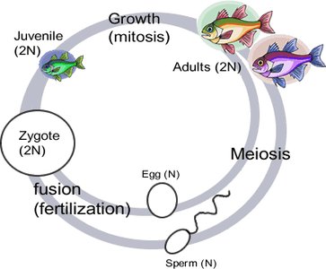
The animal kingdom, or Kingdom Animalia, comprises over 1.3 million described species, with millions more yet to be identified. Animals are eukaryotic, multicellular organisms that lack cell walls and possess an extensive extracellular matrix (ECM). They are heterotrophic, ingesting food rather than absorbing it, and most move under their own power at some stage in their life cycle. The majority reproduce sexually, with a dominant diploid stage.
Eukaryotes: Cells contain membrane-bound organelles.
Multicellular: Composed of multiple cells with specialized functions.
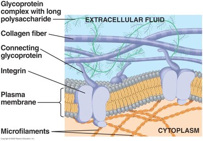
Extracellular Matrix (ECM): Provides structural support and cell signaling.
Heterotrophic: Obtain energy by consuming other organisms.
Movement: All animals move at some point in their life cycle.
Nerve and Muscle Cells: Present in all animals except sponges.
Sexual Reproduction: Most animals reproduce sexually, with gametes produced by meiosis.
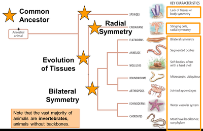
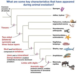
Phylogenetic Relationships and Key Innovations
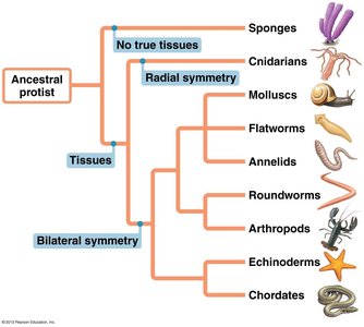
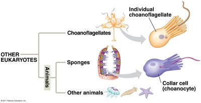
Animal evolution is marked by several key innovations, including multicellularity, embryonic tissue layers, symmetry, and specialized structures. The animal tree is rooted in a common ancestor, likely a protist similar to choanoflagellates.
Multicellularity: Originated in sponges.
Embryonic Tissue Layers: Eumetazoans possess true tissues organized into germ layers.
Symmetry: Radial symmetry (e.g., cnidarians) and bilateral symmetry (most animals).
Cephalization: Concentration of sensory organs at the anterior end.
Vertebrae, Jaws, Amniotic Egg, Limbs, Endothermy: Innovations in vertebrate evolution.
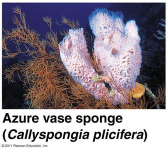
Phylum Porifera (Sponges)
Basic Biology and Structure
Sponges are basal metazoans, representing the earliest-diverging animal lineage. They are asymmetrical, lack true tissues and organs, and have a cell-level organization. Sponges are primarily marine, with some freshwater species, and are benthic (bottom-dwelling).
Choanocytes: Collar cells that create water currents and capture food via phagocytosis.
Amoebocytes: Transport nutrients, produce skeletal fibers, and differentiate into other cell types.
Mesohyl: Gelatinous matrix between cell layers.
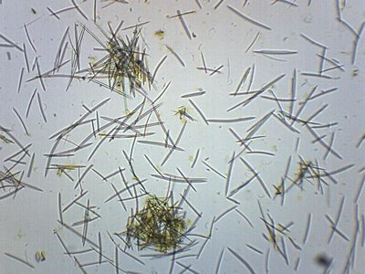
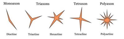
Spicules: Skeleton made of silica, calcium carbonate, or spongin fibers.
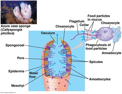
Anatomy and Canal Systems
Sponges have a sac-like body with two layers and many pores (ostia). Water enters through ostia, travels into the spongocoel (internal cavity), and exits through the osculum. The internal skeleton is made of spongin and/or spicules.
Asconoid: Simplest canal system; water flows directly from ostia to spongocoel to osculum.
Syconoid: Ostia lead into folded canals lined with choanocytes.
Leuconoid: Most complex; numerous small flagellated chambers and a network of canals.
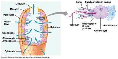
Feeding and Digestion
Sponges are suspension feeders, filtering microscopic particles from water. Choanocytes trap food and form food vacuoles, which are passed to amoebocytes for intracellular digestion. Excretion and digestion occur via diffusion.
Reproduction and Development
Asexual: Fragmentation, budding, and release of gemmules (aggregations of amoebocytes).
Sexual: Most are monoecious (produce both egg and sperm at different times). Larvae are ciliated and mobile for dispersal; adults are sessile.
Classes of Sponges
Class | Main Features |
|---|---|
Calcarea | Small marine sponges, spicules of calcium carbonate |
Demospongiae | Largest class, siliceous spicules, brightly colored |
Hexactinellidae | "Glass sponges", six-rayed siliceous spicules |
Homoscleromorpha | Fewer than 100 species, more recent addition |
Origin of Embryonic Tissue Layers
Germ Layers and Eumetazoa
Sponges lack true tissues, while other animals (Eumetazoa) possess embryonic tissues organized into germ layers. Germ layers give rise to various tissues and organs in the adult animal.
Diploblasts: Two germ layers (ectoderm and endoderm); includes cnidarians and ctenophores.
Triploblasts: Three germ layers (ectoderm, mesoderm, endoderm); includes most other animals.
Germ Layer Functions
Ectoderm: Skin, nervous system, simple muscular system.
Endoderm: Lining of digestive tract, reproductive structures.
Mesoderm (triploblasts only): Circulatory system, muscle, internal structures (bone, organs).
Symmetry in Animals
Radial vs. Bilateral Symmetry
Symmetry is a key innovation in animal evolution. Radial symmetry is found in cnidarians and ctenophores, while bilateral symmetry characterizes most other animals.
Radial Symmetry: No front/back or left/right; often sessile or planktonic.
Bilateral Symmetry: Dorsal/ventral, right/left, anterior/posterior; associated with active movement and cephalization.
Phylum Ctenophora (Comb Jellies)
Basic Biology
Ctenophores are marine, radially symmetrical diploblasts. They are pelagic swimmers, many are bioluminescent, and they lack stinging cells. Instead, they use colloblasts (adhesive cells) to capture food. Movement is achieved via cilia on comb-like plates.
Phylum Cnidaria (Jellyfish, Corals, Sea Anemones, Hydras)
Basic Biology and Structure
Cnidarians are radially symmetrical diploblasts with true tissues but no organs. They are carnivorous, capturing prey with tentacles equipped with cnidocytes (stinging cells containing nematocysts).
Nerve Net: Diffuse network for basic sensory and motor functions.
Muscle-like Cells: Enable simple movement and contraction.
Gastrovascular Cavity: Single opening for digestion and excretion.
Body Forms: Polyp (sessile) and medusa (free-swimming); many alternate between forms.
Feeding and Digestion
Carnivorous: Capture prey with tentacles.
Cnidocytes: Specialized stinging cells for prey capture and defense.
Incomplete Digestive Tract: Extracellular digestion in gastrovascular cavity, followed by intracellular digestion.
Reproduction and Life Cycles
Asexual: Budding, fission, fragmentation (especially in polyps).
Sexual: Gametes released into water; fertilization occurs externally. Medusae typically reproduce sexually.
Alternation of Forms: Many species alternate between polyp and medusa forms.
Major Cnidarian Groups
Group | Main Features |
|---|---|
Hydrozoa | Alternation of polyp/medusa; includes Hydra, Obelia, Portuguese man-of-war |
Scyphozoa | "True jellyfish"; medusa-dominant life cycle |
Cubozoa | Box jellies; highly toxic nematocysts, complex eyes |
Anthozoa | Corals and sea anemones; polyp-only, coral polyps build reefs |
Ecological Importance
Coral Reefs: Among the most productive and biodiverse ecosystems.
Reef Formation: Coral polyps + zooxanthellae (photosynthetic dinoflagellate symbionts).
Summary Table: Key Innovations in Animal Evolution
Innovation | Group | Significance |
|---|---|---|
Multicellularity | Sponges | Cell specialization, larger body size |
True Tissues | Eumetazoa | Complex body structures |
Radial Symmetry | Cnidarians, Ctenophores | Adaptation for sessile/planktonic life |
Bilateral Symmetry | Most animals | Active movement, cephalization |
Cephalization | Bilateral animals | Concentration of sensory organs |
Vertebrae, Jaws, Amniotic Egg, Limbs | Vertebrates | Predation, terrestrial adaptation |
