 Back
L 1.2: Introduction to Scientific Inquiry and Microscopy in Biology
Study Guide - Smart Notes
 Tailored notes based on your materials, expanded with key definitions, examples, and context.
L 1.2
Course Introduction: Scientific Research and the Scientific Method
Overview of Scientific Research
Scientific research is a systematic process used to answer questions and solve problems in biology. It relies on evidence, logical reasoning, and repeatable experiments to build knowledge about the natural world.
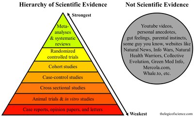
Scientific Evidence: Evidence is ranked by reliability, with meta-analyses and systematic reviews being the strongest, and case reports or opinion papers being the weakest.
Not Scientific Evidence: Anecdotes, personal beliefs, and unverified online sources do not constitute scientific evidence.
The Scientific Method
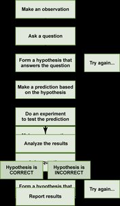
The scientific method is a logical, stepwise approach to problem-solving and experimentation in biology. It ensures that findings are based on objective, repeatable observations.
Steps: Observation, question, hypothesis formation, prediction, experimentation, analysis, and conclusion.
Hypothesis: A testable explanation for an observation or scientific problem.
Experiment: A controlled procedure to test the hypothesis.
Analysis: Data are interpreted to determine if the hypothesis is supported or refuted.
Types of Research: Fundamental vs. Applied
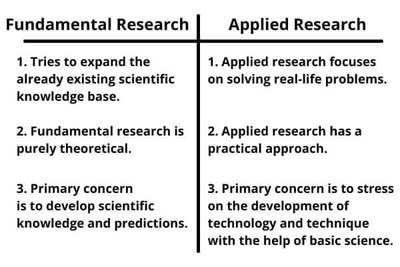
Biological research can be classified as fundamental (basic) or applied, depending on its goals and methods.
Fundamental Research | Applied Research |
|---|---|
Expands scientific knowledge base | Solves real-life problems |
Theoretical focus | Practical approach |
Develops predictions and understanding | Develops technology and techniques |
Microscopy: Tools for Observing Life
History of Microscopy
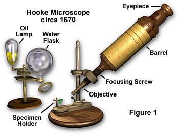
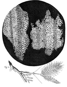
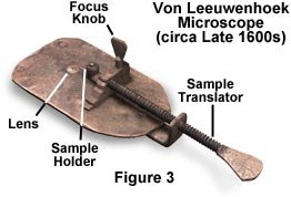
Microscopes revolutionized biology by allowing scientists to observe cells and microorganisms. Key historical figures include Robert Hooke and Antonie van Leeuwenhoek.
Robert Hooke: Discovered cork cells and coined the term "cell." Used a compound microscope.
Antonie van Leeuwenhoek: Built simple microscopes, observed living cells such as bacteria and sperm, and improved lens-making techniques.
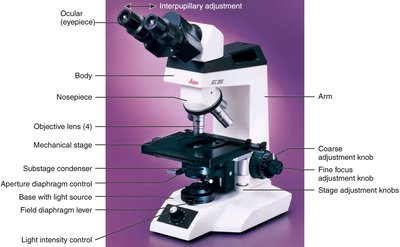
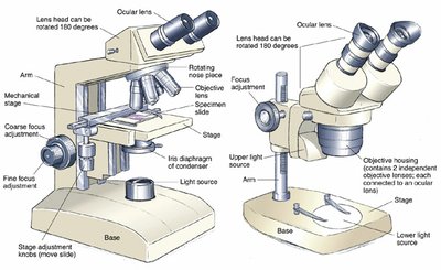
Parts and Handling of the Compound Microscope
Proper use and care of the microscope are essential for accurate observations.
Key Parts: Ocular lens (eyepiece), objective lenses, stage, coarse and fine focus knobs, light source.
Handling: Carry with one hand on the arm and one under the base. Clean lenses before and after use.
Focusing: Start with low power, then switch to higher power. Parfocal microscopes maintain focus when changing objectives.
Oil Immersion: Use oil only with the oil immersion lens to increase resolution.

Principles of Magnification and Image Formation
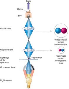
Microscopes use lenses to magnify specimens and form images. Magnification is the product of the objective and ocular lens powers.
Magnification:
Image Formation: The objective lens forms a real image, which is further magnified by the ocular lens to produce a virtual image.
Photographic Magnification: If a 10 µm object appears as 10 cm in a photo, the magnification is 10,000X.
Orientation of Images
Microscope images appear inverted and reversed due to the optics of the lenses.
Moving the slide left makes the image move right, and vice versa.

Types of Microscopes
Different microscopes are used for various applications in biology.
Bright-field: Most common; requires stained specimens for contrast.
Dark-field: Illuminates specimens against a dark background; useful for live, unstained samples.
Phase-contrast: Enhances contrast in unstained cells by amplifying differences in light phase.
DIC (Nomarski): Provides high-contrast, detailed images of transparent specimens.
Dissecting/Stereo Microscope: Used for low-magnification observation of larger specimens.
Resolving Power and Numerical Aperture
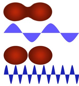
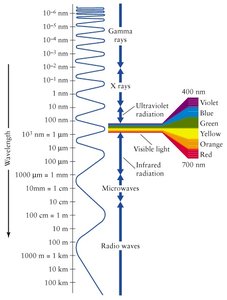
Resolution is the ability to distinguish two points as separate. It depends on the wavelength of light and the numerical aperture (NA) of the lens.
Resolution Formula: , where is the minimum resolvable distance, is the wavelength, and is numerical aperture.
Numerical Aperture: A measure of a lens's ability to gather light and resolve fine specimen detail at a fixed object distance.
Oil Immersion: Increases NA by reducing light refraction, improving resolution.
Microscopy Techniques: Polarizing, Fluorescence, and Confocal
Advanced microscopy techniques enhance visualization of specific structures or molecules.
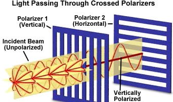
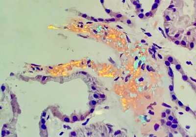
Polarizing Microscopy: Uses polarized light to detect structures with specific optical properties, such as crystals or amyloid deposits.
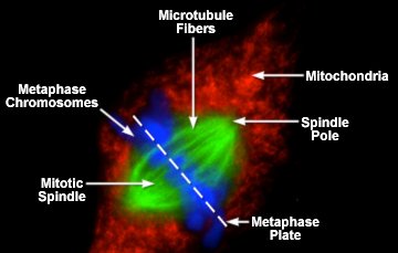
Fluorescence Microscopy: Uses fluorescent dyes and UV light to visualize specific cell components.
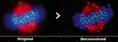
Confocal Microscopy: Uses point illumination and spatial pinholes to create sharp, 3D images of thick specimens.
Electron Microscopy
Electron microscopes use electron beams instead of light, allowing much higher resolution and magnification.
Transmission Electron Microscope (TEM): Electrons pass through thin specimens, revealing internal structures at high resolution.
Scanning Electron Microscope (SEM): Electrons scan the specimen surface, producing detailed 3D images of surfaces.
Magnification: Ranges from 5,000X to 1,000,000X.
Photography in Biology
Microscopy Photography
Photographs taken through microscopes are essential for documenting observations and sharing results in biology. The magnification of the photograph is the product of the microscope magnification and the enlargement factor of the print.
Applications: Used in teaching, research, and publication to illustrate cellular structures and processes.
Practice Questions and Applications
Identify the type of microscope used to view an Elodea leaf.
Relate observed structures to their biological function.
Interpret images and videos of mitosis and other cellular processes.
Additional info: This guide covers foundational concepts from Ch. 1 (Scientific Inquiry), Ch. 6 (A Tour of the Cell), and Ch. 7 (Membrane Structure and Function), as well as microscopy techniques relevant to cell biology.
