 Back
Labeling Animal Cell Mitosis Phases in Microscopy Images
Study Guide - Smart Notes
 Tailored notes based on your materials, expanded with key definitions, examples, and context.
Q: Label the diagrams of animal cell mitosis using the terms: Interphase, Prophase, Metaphase, Anaphase, Telophase/Cytokinesis.
Background
Topic: Cell Cycle and Mitosis
This question tests your ability to recognize and distinguish the different phases of mitosis in animal cells as seen under a microscope. Understanding these phases is fundamental for studying how cells divide and ensure genetic continuity.
Key Terms and Concepts:
Interphase: The cell is not dividing; DNA is in the form of chromatin and the nucleus is intact.
Prophase: Chromosomes condense and become visible, spindle fibers begin to form, and the nuclear envelope breaks down.
Metaphase: Chromosomes align at the cell's equatorial (metaphase) plate.
Anaphase: Sister chromatids separate and move toward opposite poles of the cell.
Telophase/Cytokinesis: Chromosomes decondense, nuclear envelopes reform, and the cytoplasm divides (cleavage furrow forms in animal cells).
Step-by-Step Guidance
Examine each cell in the images and look for key features: Is the DNA diffuse (chromatin) or condensed (chromosomes)? Is the nuclear envelope present or absent?
Identify Interphase cells by their intact nucleus and uncondensed chromatin. These cells will not show visible chromosomes.
Look for Prophase cells where chromosomes start to condense and become visible, but are not yet aligned in the center. The nuclear envelope may be breaking down.
Find Metaphase cells by locating those with chromosomes lined up along the center (metaphase plate) of the cell.
Spot Anaphase cells by identifying those where sister chromatids are being pulled apart toward opposite poles.
Identify Telophase/Cytokinesis by looking for cells where chromosomes are at the poles, nuclear envelopes are reforming, and the cell may be pinching in two (cleavage furrow).
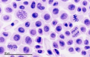
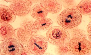
Try labeling each phase in the images before checking the answer!
Final Answer:
Typical order for labeling (left to right/top to bottom):
Interphase: Nucleus intact, chromatin not condensed.
Prophase: Chromosomes condensing, spindle forming, nuclear envelope breaking down.
Metaphase: Chromosomes aligned at equator/metaphase plate.
Anaphase: Sister chromatids separating and moving to opposite poles.
Telophase/Cytokinesis: Nuclear envelopes reforming, chromosomes decondensing, cytoplasm dividing.
Each image contains cells in various stages—look for these features to match the correct phase to each cell.
