 Back
Large Biological Molecules: Proteins, Carbohydrates, Lipids, and Nucleic Acids
Study Guide - Smart Notes
 Tailored notes based on your materials, expanded with key definitions, examples, and context.
Large Biological Molecules – Proteins
Protein Structure and Function
Proteins are essential macromolecules composed of amino acid monomers. Their structure determines their diverse functions in biological systems.
Protein: A functional biological molecule consisting of one or more polypeptides folded into a specific three-dimensional structure.
Polypeptide: A polymer of amino acids linked by peptide bonds.
Peptide bond: A covalent bond formed between the carboxyl group of one amino acid and the amino group of another, releasing water (dehydration synthesis).
Amino acid: The monomer unit of proteins, each containing a central (α) carbon, an amino group, a carboxyl group, a hydrogen atom, and a variable R group (side chain).
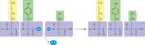
Conformation: The three-dimensional shape of a protein, critical for its function.
Denaturation: The loss of a protein's native structure due to external stress (e.g., heat, pH, chemicals), resulting in loss of function.
Levels of Protein Structure
Proteins have four levels of structure, each contributing to their final shape and function:
Level | Explanation |
|---|---|
Primary | Linear sequence of amino acids in a polypeptide, held together by peptide bonds. |
Secondary | Regular folding of the polypeptide backbone into α helices and β pleated sheets, stabilized by hydrogen bonds between backbone atoms. |
Tertiary | Overall three-dimensional shape formed by interactions between side chains (R groups), including hydrophobic interactions, van der Waals forces, hydrogen bonds, ionic bonds, and disulfide bridges. |
Quaternary | Association of two or more polypeptide chains into a functional protein complex. |
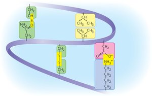
Hydrophobic effects: Nonpolar side chains cluster away from water, stabilizing the protein's core.
Van der Waals forces: Weak attractions between nonpolar side chains.
Disulfide bridge: Covalent bond between sulfur atoms of two cysteine residues, reinforcing structure.
Chaperone proteins: Assist in the proper folding of other proteins, preventing misfolding and aggregation.
Example: Enzymes are globular proteins with complex tertiary and sometimes quaternary structures, enabling them to catalyze biochemical reactions.
Large Biological Molecules – Carbohydrates
Carbohydrate Structure and Function
Carbohydrates are sugars and their polymers, serving as energy sources and structural materials.
Monosaccharide: Simple sugar monomer (e.g., glucose, fructose).
Disaccharide: Two monosaccharides joined by a glycosidic bond (e.g., sucrose, lactose, maltose).
Polysaccharide: Long chains of monosaccharides (e.g., starch, glycogen, cellulose).
Glycosidic bond: Covalent bond joining two monosaccharides via dehydration synthesis.
Monosaccharide formula examples:
Hexose (6C):
Pentose (5C):
Triose (3C):
All sugars contain a carbonyl group (C=O) and multiple hydroxyl groups (–OH).
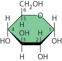
Example: Glucose is a hexose sugar with six carbons, each corner of the ring (except the oxygen) represents a carbon atom.
Polysaccharide Types and Functions
Type | Examples (Plants) | Examples (Animals) |
|---|---|---|
Energy storage | Starch | Glycogen |
Structural building block | Cellulose | Chitin (in fungi and some animals) |
Starch and glycogen are branched polymers, making them efficient for energy storage. Cellulose is unbranched, providing structural support in plant cell walls.
Large Biological Molecules – Lipids
Lipid Structure and Function
Lipids are hydrophobic molecules, including fats, phospholipids, and steroids. They are not true polymers but are assembled from smaller components.
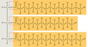
Fat: Composed of glycerol and three fatty acids (triacylglycerol), joined by ester linkages.
Saturated fatty acid: No double bonds; straight chains; solid at room temperature (animal fats).
Unsaturated fatty acid: One or more double bonds; kinked chains; liquid at room temperature (plant oils).
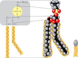
Phospholipid: Glycerol, two fatty acids, and a phosphate group; forms the bilayer of cell membranes.
Bilayer: Arrangement of phospholipids in membranes, with hydrophilic heads facing water and hydrophobic tails inward.
Steroid: Lipid with four fused rings; includes cholesterol and hormones like testosterone and estrogen.
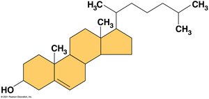
Example: Cholesterol stabilizes animal cell membranes; steroid hormones regulate physiology.
Large Biological Molecules – Nucleic Acids
Nucleic Acid Structure and Function
Nucleic acids (DNA and RNA) store and transmit genetic information. Their monomers are nucleotides, each consisting of a phosphate group, a five-carbon sugar (deoxyribose or ribose), and a nitrogenous base.
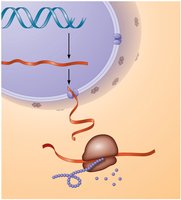
DNA: Deoxyribonucleic acid; double helix; stores genetic information.
RNA: Ribonucleic acid; single-stranded; involved in gene expression.
Nucleotide: Monomer of nucleic acids; composed of a phosphate, a pentose sugar, and a nitrogenous base.
Pyrimidines: Single-ring bases (cytosine, thymine, uracil).
Purines: Double-ring bases (adenine, guanine).
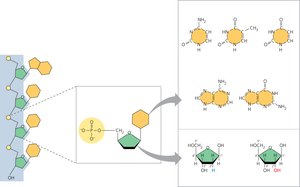
DNA bases: Adenine (A), Thymine (T), Cytosine (C), Guanine (G)
RNA bases: Adenine (A), Uracil (U), Cytosine (C), Guanine (G)
Difference between ribose and deoxyribose: Deoxyribose lacks one oxygen atom compared to ribose.
DNA Structure
DNA consists of two antiparallel strands forming a double helix. The backbone is made of alternating sugar and phosphate groups, while the rungs are pairs of nitrogenous bases connected by hydrogen bonds.
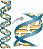
Base pairing: Adenine pairs with thymine (2 hydrogen bonds), cytosine pairs with guanine (3 hydrogen bonds).
Example: For the sequence TAGGCCT, the complementary strand is ATCCGGA.
Equation for dehydration synthesis (joining two monosaccharides):
Additional info: The loss of water explains why the disaccharide does not follow the 1:2:1 ratio of C:H:O seen in monosaccharides.
