 Back
Lipids, Membranes, and Membrane Transport
Study Guide - Smart Notes
 Tailored notes based on your materials, expanded with key definitions, examples, and context.
Ch 6 – Lipids and Membranes
Overview of Lipids
Lipids are a diverse group of hydrophobic biological macromolecules that play critical roles in energy storage, membrane structure, and signaling. The three main types of lipids are steroids, fats (triglycerides), and phospholipids. All lipids are united by their insolubility in water due to a high proportion of nonpolar C–H bonds.
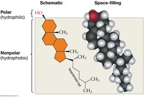
Steroids: Characterized by a four-ring structure. Function as hormones and membrane components (e.g., cholesterol).
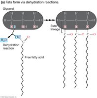
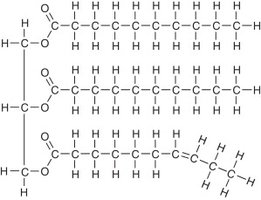
Fats (Triglycerides): Composed of three fatty acids linked to a glycerol backbone. Serve as energy storage molecules.
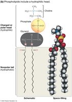
Phospholipids: Consist of a glycerol backbone, two fatty acid tails, and a phosphate-containing head group. Major structural component of cell membranes.
What unites lipids? All lipids are primarily hydrophobic due to their long hydrocarbon chains or rings.
Types of Lipids
Steroids
Steroids have a core structure of four fused hydrocarbon rings with various functional groups attached. Cholesterol is a common steroid in animal cell membranes, contributing to membrane fluidity and serving as a precursor for steroid hormones.
Fats (Triglycerides)
Fats are formed by dehydration reactions between glycerol and three fatty acids, resulting in ester linkages. They are used for long-term energy storage and insulation in animals.
Phospholipids
Phospholipids are amphipathic molecules, meaning they have both hydrophilic (polar) and hydrophobic (nonpolar) regions. The hydrophilic head contains a phosphate group, while the hydrophobic tails are fatty acid chains. This structure is essential for forming biological membranes.
Saturation of Fatty Acid Tails
Saturated vs. Unsaturated Fatty Acids
The degree of saturation in fatty acid tails affects the physical properties of lipids:
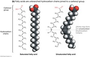
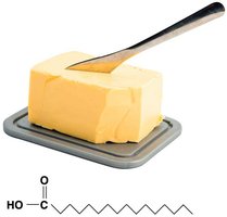
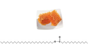
Saturated fatty acids: Contain only single bonds between carbon atoms, resulting in straight chains that pack tightly together. Typically solid at room temperature (e.g., butter, beeswax).
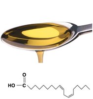
Unsaturated fatty acids: Contain one or more double bonds, introducing kinks that prevent tight packing. Usually liquid at room temperature (e.g., oils).
Physical Properties of Fats and Oils
The presence of double bonds (unsaturation) lowers the melting point of fats, making them liquid at room temperature. Saturated fats are solid due to their straight chains and tight packing.
Butter: High in saturated fats, solid at room temperature.
Beeswax: Highly saturated, very solid.
Safflower oil: High in unsaturated fats, liquid at room temperature.
Hydrogenated oil: Unsaturated fats that have been artificially saturated, making them solid.
Lipid Organization in Water
Micelles and Bilayers
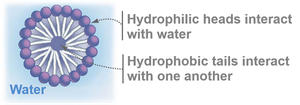
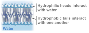
When placed in water, amphipathic lipids spontaneously organize to minimize free energy:
Micelles: Spherical structures formed by single-chain lipids (e.g., fatty acids) with hydrophobic tails inward and hydrophilic heads outward.
Lipid bilayers: Double-layered sheets formed by phospholipids, with hydrophobic tails sandwiched between hydrophilic heads facing the aqueous environment.
Lipid Bilayers and Membrane Permeability
Selectively Permeable Membranes
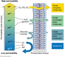
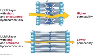
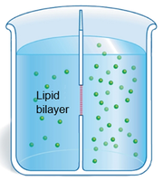
Lipid bilayers are selectively permeable, allowing some substances to cross more easily than others. Small, nonpolar molecules (e.g., O2, CO2) diffuse rapidly, while ions and large polar molecules are largely excluded.
Factors Affecting Membrane Permeability
Fatty acid tail length: Shorter tails increase permeability; longer tails decrease it.
Degree of saturation: Unsaturated tails (with kinks) increase permeability; saturated tails decrease it.
Cholesterol content: Cholesterol reduces membrane permeability by filling spaces between phospholipids.
Diffusion and Osmosis
Diffusion
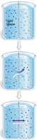
Diffusion is the movement of molecules from an area of higher concentration to an area of lower concentration, driven by the concentration gradient, until equilibrium is reached.
Osmosis
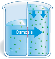
Osmosis is the diffusion of water across a selectively permeable membrane. Water moves from regions of low solute concentration to regions of high solute concentration to equalize solute concentrations on both sides of the membrane.
Structure of the Cell Membrane: Fluid-Mosaic Model
Fluid-Mosaic Model
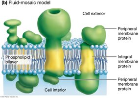
The cell membrane is described as a fluid-mosaic because it is composed of a dynamic, fluid phospholipid bilayer with proteins embedded throughout. The 'fluid' aspect refers to the lateral movement of lipids and proteins, while 'mosaic' refers to the patchwork of proteins that float in or on the lipid bilayer.
Membrane Proteins and Transport
Types of Membrane Transport Proteins
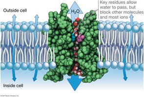
Channel proteins: Form pores that allow specific ions or water molecules to pass through the membrane by facilitated diffusion.
Carrier proteins: Undergo conformational changes to transport specific molecules across the membrane.
Pumps: Use energy (usually from ATP) to move substances against their concentration gradients (active transport).
Cotransporters: Couple the movement of one molecule with its gradient to the movement of another molecule against its gradient (secondary active transport).
Examples of Channel Proteins
Aquaporins: Water channel proteins that facilitate rapid water movement across membranes.
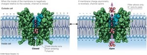
Voltage-gated K+ channels: Open or close in response to changes in membrane potential, allowing selective passage of potassium ions.
Example of a Carrier Protein – GLUT-1
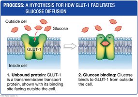
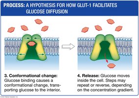
GLUT-1 is a glucose transporter that facilitates the diffusion of glucose across the plasma membrane. It binds glucose, undergoes a conformational change, and releases glucose on the other side of the membrane.
Active Transport – Sodium-Potassium Pump
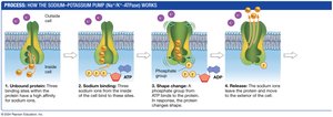
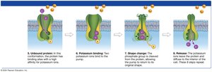
The sodium-potassium pump (Na+/K+-ATPase) is an example of primary active transport. It uses ATP to move 3 Na+ ions out of the cell and 2 K+ ions into the cell, establishing an electrochemical gradient essential for many cellular processes.
Secondary Active Transport (Cotransport)
Secondary active transport uses the energy stored in an electrochemical gradient (often established by the Na+/K+ pump) to drive the transport of other molecules against their gradients. Cotransporters move one molecule with its gradient and another against its gradient. For example, the sodium-glucose cotransporter (SGLT) uses the Na+ gradient to import glucose into cells.
Example: H+/sucrose cotransporter (SUT1) in plants uses a proton gradient to import sucrose.
Additional info: This guide covers the structure and function of lipids, the organization of biological membranes, and the mechanisms of membrane transport, as outlined in General Biology Chapter 6.
