 Back
Meiosis, Karyotyping, and Mutations: A Study Guide
Study Guide - Smart Notes
 Tailored notes based on your materials, expanded with key definitions, examples, and context.
Meiosis, Karyotyping, and Mutations
Important Terms and Concepts
This section introduces the foundational vocabulary and concepts necessary to understand meiosis, karyotyping, and mutations in the context of genetics and cell biology.
Asexual reproduction: The creation of genetically identical offspring by a single parent, without the participation of gametes (sperm and egg).
Sexual reproduction: A biological process involving the joining of gametes, typically from two parents, resulting in genetically unique offspring.
Meiosis: A specialized form of cell division in gonads to produce gametes, reducing the chromosome number by half.
Somatic cell: Any body cell except gametes; in humans, these contain 46 chromosomes.
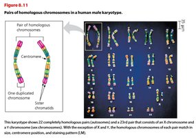
Karyotype: An organized profile of an organism’s chromosomes, arranged by size and centromere position, used to detect chromosomal abnormalities.
Homologous chromosomes: Chromosome pairs of the same length, centromere position, and gene loci, one inherited from each parent.
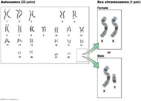
Sex chromosomes: Chromosomes that determine an individual's sex (XX for female, XY for male in humans).
Autosomes: Non-sex chromosomes; humans have 44 autosomes (22 pairs).
Diploid (2n): Cells containing two sets of chromosomes (pairs of homologous chromosomes).
Haploid (n): Cells with a single set of chromosomes, such as gametes.
Homologous Chromosomes and Karyotyping
Homologous chromosomes are central to understanding inheritance and genetic variation. Karyotyping is a laboratory technique used to visualize and analyze the chromosomal composition of an individual.
Humans have 23 pairs of chromosomes: 22 pairs of autosomes and 1 pair of sex chromosomes.
In females, both sex chromosomes are X; in males, one is X and the other is Y.
Karyotypes are used to detect chromosomal abnormalities, such as extra or missing chromosomes.
Gametes and the Human Life Cycle
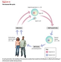
The human life cycle involves alternating diploid and haploid stages, with meiosis and fertilization ensuring genetic continuity and variation.
Humans are diploid organisms; somatic cells have 46 chromosomes (2n).
Gametes (sperm and egg) are haploid (n = 23), produced by meiosis.
Fertilization restores the diploid number, forming a zygote (2n = 46).
Mitotic divisions ensure all somatic cells inherit the correct chromosome number.

How Meiosis Halves Chromosome Number
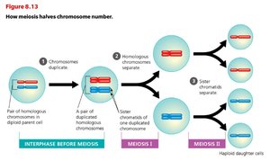
Meiosis is a two-stage division process that reduces the chromosome number by half, producing four genetically unique haploid cells from one diploid cell.
Meiosis I separates homologous chromosomes.
Meiosis II separates sister chromatids.
Crossing over during Prophase I increases genetic diversity.
The Process of Meiosis
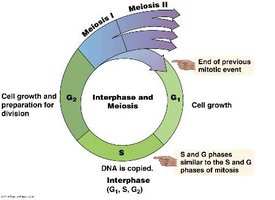
Meiosis consists of two sequential divisions: Meiosis I and Meiosis II, each with distinct phases.
Interphase: DNA is duplicated during the S phase.
Meiosis I: Homologous chromosomes are separated into two cells.
Meiosis II: Sister chromatids are separated, resulting in four haploid cells.
Crossing over: Exchange of genetic material between non-sister chromatids during Prophase I.
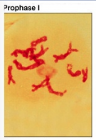
Meiosis I: Prophase I
Nuclear envelope breaks down, microtubules assemble, chromosomes condense.
Crossing over occurs, increasing genetic variation.

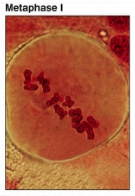
Meiosis I: Metaphase I
Homologous pairs align at the cell equator.
Random alignment leads to genetic variation in gametes.
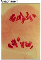
Meiosis I: Anaphase I, Telophase I, and Cytokinesis
Anaphase I: Homologous chromosomes are separated by shortening microtubules.
Telophase I: Nuclear envelope reforms around chromosomes.
Cytokinesis: Two haploid daughter cells are formed.
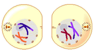
When Meiosis Goes Wrong: Non-disjunction
Non-disjunction is an error in meiosis where chromosomes fail to separate properly, leading to gametes with abnormal chromosome numbers.
Can occur during Meiosis I or II.
Results in disorders such as Down syndrome (Trisomy 21).
Karyotype Analysis and Chromosomal Disorders
Karyotyping is used to detect chromosomal abnormalities, such as extra or missing chromosomes, which can lead to genetic disorders.
Down syndrome (Trisomy 21): Presence of an extra chromosome 21 (47 chromosomes total).
Abnormal numbers of sex chromosomes: Can result in conditions such as Turner syndrome (XO) or Klinefelter syndrome (XXY).
Mutations
Mutations are changes to the genetic information of a cell or virus. They can affect single nucleotides or larger chromosomal segments, and are a source of genetic diversity and disease.
Mutation: Any change to the DNA sequence.
Some diseases, such as sickle-cell anemia, are caused by a single nucleotide change.
Types of Mutations
Nucleotide substitutions: Replacement of one nucleotide pair with another.
Nucleotide insertions or deletions: Addition or loss of nucleotide pairs, which can cause frameshift mutations.
Substitution Mutations
Silent mutation: Alters a codon but does not change the amino acid.
Missense mutation: Changes one amino acid in the protein.
Nonsense mutation: Changes a codon to a stop codon, resulting in a truncated protein.
Frameshift Mutations
Caused by insertions or deletions that shift the reading frame of the genetic message.
Can drastically alter the resulting protein.
Mutagens
Mutagen: A chemical or physical agent that causes mutations.
Physical mutagens: X-rays, ultraviolet (UV) light.
Chemical mutagens: Cigarette smoke, benzoyl peroxide.
