 Back
BackMeiosis: Mechanisms and Genetic Variation
Study Guide - Smart Notes
 Tailored notes based on your materials, expanded with key definitions, examples, and context.
Chapter 13: Meiosis
Introduction to Meiosis and Genetics
Meiosis is a specialized form of cell division that reduces the chromosome number by half, producing haploid gametes from diploid cells. This process is fundamental to sexual reproduction and is a key driver of genetic diversity in eukaryotes. Genetics, the study of inheritance, explores how traits are transmitted from one generation to the next and how variation arises.
Inheritance: The transmission of traits from parents to offspring.
Genetics: The scientific study of inherited similarities and variation.
Genes: Units of heredity, composed of DNA sequences, that code for proteins and determine traits.
Gametes: Haploid reproductive cells (sperm and egg) that carry genes to the next generation.
Somatic cells: All body cells except gametes; these are diploid.
Chromosomes and Genetic Variation
Chromosome Structure and Homologous Pairs
Chromosomes are long DNA molecules containing many genes. In diploid organisms, chromosomes exist in pairs called homologous chromosomes, which carry genes for the same traits but may have different alleles.
Diploid (2n): Cells with two sets of chromosomes (e.g., human somatic cells have 46 chromosomes).
Haploid (n): Cells with one set of chromosomes (e.g., human gametes have 23 chromosomes).
Locus (plural: loci): The specific location of a gene on a chromosome.
Homologous chromosomes: Chromosome pairs, one from each parent, that are similar in length, gene position, and centromere location.
One extra or missing chromosome can cause genetic disorders, highlighting the importance of correct chromosome number.
Overview of Meiosis
Purpose and Process
Meiosis consists of two sequential divisions (Meiosis I and Meiosis II) but only one round of DNA replication. The result is four haploid cells, each genetically distinct from the parent cell and from each other.
Meiosis I: Homologous chromosomes separate.
Meiosis II: Sister chromatids separate (similar to mitosis).
Phases of Meiosis I
Prophase I
Homologous chromosomes pair up and exchange genetic material through crossing over at structures called chiasmata. The nuclear envelope fragments, and spindle fibers begin to form.
Crossing over: Exchange of genetic material between non-sister chromatids, increasing genetic variation.
Chiasmata: X-shaped regions where crossing over occurs.

Metaphase I
Pairs of homologous chromosomes align at the metaphase plate. Microtubules from opposite poles attach to the kinetochores of homologous chromosomes.

Anaphase I
Homologous chromosomes are pulled to opposite poles of the cell, while sister chromatids remain attached at their centromeres.

Telophase I and Cytokinesis
Each half of the cell now has a haploid set of chromosomes, each still consisting of two sister chromatids. Cytokinesis divides the cytoplasm, forming two haploid cells.
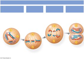
Summary of Meiosis I
Meiosis I separates homologous chromosomes, reducing the chromosome number by half. The resulting cells are haploid but each chromosome still consists of two sister chromatids.

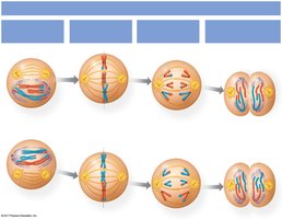
Phases of Meiosis II
Overview
Meiosis II resembles mitosis, separating sister chromatids without further DNA replication. It consists of four phases: Prophase II, Metaphase II, Anaphase II, and Telophase II with cytokinesis.
Prophase II
Spindle apparatus forms in each haploid cell, and chromosomes move toward the metaphase II plate.

Metaphase II
Chromosomes align individually along the metaphase plate. Microtubules attach to kinetochores of sister chromatids.

Anaphase II
Sister chromatids are finally separated and pulled to opposite poles of the cell.

Telophase II and Cytokinesis
Chromosomes arrive at the poles, nuclei reform, and chromosomes decondense. Cytokinesis produces four genetically unique haploid cells, each with 23 chromosomes in humans.
Genetic Variation in Sexual Reproduction
Mechanisms of Variation
Crossing Over: Occurs during Prophase I, exchanging genetic material between homologous chromosomes.
Independent Assortment: Random orientation of homologous pairs during Metaphase I leads to different combinations of maternal and paternal chromosomes in gametes.
Fertilization: Random fusion of gametes further increases genetic diversity.
Comparison: Mitosis vs. Meiosis
Key Differences
Feature | Mitosis | Meiosis |
|---|---|---|
Number of divisions | 1 | 2 |
Number of daughter cells | 2 | 4 |
Chromosome number in daughter cells | Diploid (2n) | Haploid (n) |
Genetic identity | Identical to parent | Genetically unique |
Role | Growth, repair | Gamete production |
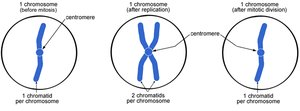
Review: Chromosome and Chromatid Numbers
Changes in Chromosome and Chromatid Number
During meiosis, cells transition from having two pairs of each chromosome (diploid) to one of each chromosome (haploid). The number of chromatids per chromosome changes with each phase:
Stage | Chromosomes per cell | Chromatids per chromosome |
|---|---|---|
Before DNA replication | 2n | 1 |
After DNA replication (before meiosis I) | 2n | 2 |
After meiosis I | n | 2 |
After meiosis II | n | 1 |
Summary
Meiosis produces four genetically unique haploid cells from one diploid cell.
Genetic variation arises from crossing over, independent assortment, and fertilization.
Meiosis is essential for sexual reproduction and maintaining chromosome number across generations.
