 Back
Meiosis: Mechanisms, Phases, and Biological Significance
Study Guide - Smart Notes
 Tailored notes based on your materials, expanded with key definitions, examples, and context.
Meiosis and Heredity
Introduction to Heredity and Genetics
Heredity is the process by which traits are transmitted from one generation to the next. Genetics is the scientific study of heredity and variation in organisms. The fundamental units of heredity are genes, which are specific sequences of DNA located at particular positions, or loci, on chromosomes. Alternative forms of a gene are called alleles.
Gene: A segment of DNA that encodes information for a specific trait.
Locus: The fixed position of a gene on a chromosome.
Allele: Different versions of a gene found at the same locus.
Chromosomes and Karyotypes
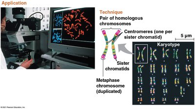
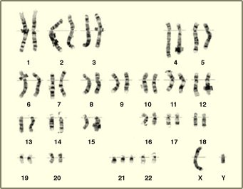
Most DNA in eukaryotic cells is organized into chromosomes. Human somatic cells contain 23 pairs of chromosomes, including 22 pairs of autosomes and one pair of sex chromosomes. Homologous chromosomes are pairs that have the same length, centromere position, and gene loci, but may carry different alleles.
Homologous chromosomes: Chromosome pairs, one from each parent, carrying genes for the same traits.
Karyotype: An ordered visual display of all chromosomes in a cell, used to detect chromosomal abnormalities.
Chromosome Number and Cell Types
Diploid and Haploid Cells
Cells can be classified based on their chromosome number:
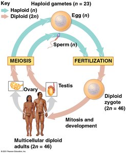
Diploid (2n): Cells with two sets of chromosomes (e.g., human somatic cells, 2n = 46).
Haploid (n): Cells with one set of chromosomes (e.g., human gametes, n = 23).
Gametes (sperm and egg) are produced by meiosis and are haploid. Fertilization restores the diploid number in the zygote, and mitosis ensures growth and development of the multicellular organism.
Overview of Meiosis
Purpose and General Features
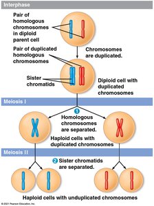
Meiosis is a specialized type of cell division that reduces the chromosome number by half, producing four genetically distinct haploid cells from one diploid cell. It consists of two sequential divisions: meiosis I (reductional division) and meiosis II (equational division).
Meiosis I: Homologous chromosomes separate, reducing chromosome number by half.
Meiosis II: Sister chromatids separate, similar to mitosis.
Phases of Meiosis
Meiosis I: Reductional Division
Meiosis I separates homologous chromosomes and consists of four main phases:
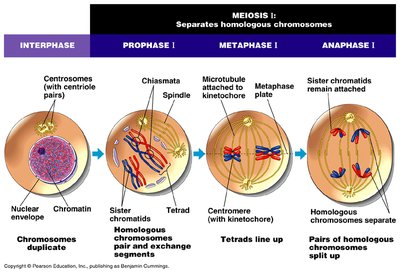
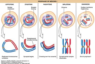
Prophase I: Chromosomes condense, homologous chromosomes pair (synapsis), and crossing over occurs at chiasmata. This phase is subdivided into leptotene, zygotene, pachytene, diplotene, and diakinesis.
Metaphase I: Homologous pairs align at the metaphase plate; microtubules attach to kinetochores.
Anaphase I: Homologous chromosomes separate and move to opposite poles; sister chromatids remain attached.
Telophase I and Cytokinesis: Two haploid cells form, each with duplicated chromosomes.
Subphases of Prophase I
Leptotene: Chromosomes start to condense.
Zygotene: Synapsis begins; homologous chromosomes pair up.
Pachytene: Crossing over occurs between non-sister chromatids.
Diplotene: Synaptonemal complex dissolves; chiasmata become visible.
Diakinesis: Chromosomes further condense; nuclear envelope fragments.
Key Structures in Prophase I
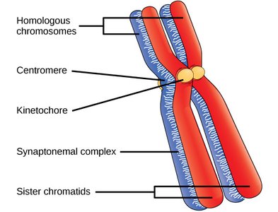
Synaptonemal complex: Protein structure that forms between homologous chromosomes during synapsis, facilitating crossing over.
Chiasmata: X-shaped regions where crossing over has occurred.
Crossing Over
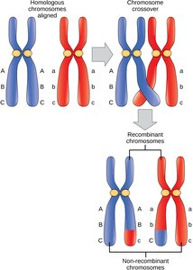
Crossing over is the exchange of genetic material between non-sister chromatids of homologous chromosomes, increasing genetic diversity in gametes.
Meiosis II: Equational Division
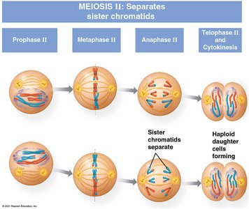
Meiosis II resembles mitosis and separates sister chromatids. It consists of:
Prophase II: Spindle apparatus forms; chromosomes move toward the metaphase plate.
Metaphase II: Chromosomes align at the metaphase plate; kinetochores attach to microtubules from opposite poles.
Anaphase II: Sister chromatids separate and move to opposite poles.
Telophase II and Cytokinesis: Chromosomes decondense, nuclei reform, and four genetically distinct haploid cells are produced.
Genetic Consequences of Meiosis
Genetic Variation
Independent assortment: Random orientation of homologous pairs during metaphase I leads to genetic variation.
Crossing over: Produces recombinant chromosomes with new allele combinations.
Karyotypes and Chromosomal Disorders
Karyotype Analysis
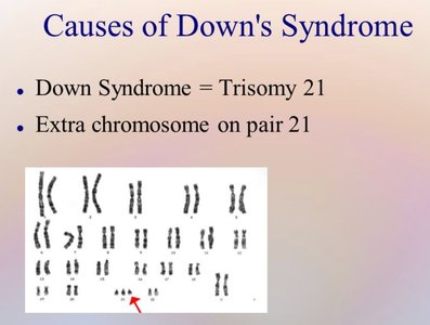
Karyotyping is used to detect chromosomal abnormalities, such as aneuploidies. Down syndrome (trisomy 21) is caused by the presence of an extra chromosome 21, often due to nondisjunction during meiosis.
Comparison of Mitosis and Meiosis
Key Differences
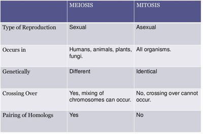
Mitosis and meiosis are both forms of cell division, but they differ in their outcomes and biological roles. The following table summarizes the main differences:
Meiosis | Mitosis | |
|---|---|---|
Type of Reproduction | Sexual | Asexual |
Occurs in | Humans, animals, plants, fungi | All organisms |
Genetically | Different | Identical |
Crossing Over | Yes, mixing of chromosomes can occur | No, crossing over cannot occur |
Pairing of Homologs | Yes | No |
Key Terms and Definitions
Heredity: Transmission of traits from one generation to the next.
Genetics: Study of heredity and variation.
Gene: Unit of heredity, a segment of DNA.
Locus: Specific location of a gene on a chromosome.
Allele: Alternative form of a gene.
Diploid (2n): Cell with two sets of chromosomes.
Haploid (n): Cell with one set of chromosomes.
Synapsis: Pairing of homologous chromosomes during prophase I of meiosis.
Crossing over: Exchange of genetic material between non-sister chromatids.
Chiasmata: Sites where crossing over occurs.
Karyotype: Ordered display of chromosomes in a cell.
Self-Review Questions
Define heredity, genetics, genes, locus, and alleles.
Define diploid and haploid cell.
What are the phases of meiosis?
Describe briefly the phases of meiosis and the events during these phases.
What is synapsis? What is crossing over? Define chiasmata.
Define karyotype. What is the cause of Down syndrome?
What are the differences between mitosis and meiosis cell division?
