 Back
Membrane Structure and Function, Cell Structure, and Protein Organization
Study Guide - Smart Notes
 Tailored notes based on your materials, expanded with key definitions, examples, and context.
Cell Structure: Animal and Plant Cells
Overview of Eukaryotic Cell Structure
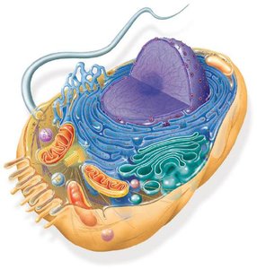
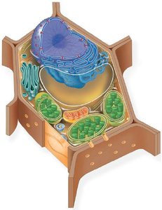
Eukaryotic cells are highly organized and contain membrane-bound organelles that perform specialized functions. Animal and plant cells share many structures but also have unique features that distinguish them.
Nucleus: Contains genetic material (DNA) and controls cellular activities.
Endoplasmic Reticulum (ER): Rough ER is studded with ribosomes and synthesizes proteins; smooth ER synthesizes lipids and detoxifies chemicals.
Golgi Apparatus: Modifies, sorts, and packages proteins and lipids for secretion or delivery to other organelles.
Mitochondria: Site of cellular respiration and ATP production.
Lysosomes: Contain digestive enzymes to break down waste (mainly in animal cells).
Cytoskeleton: Provides structural support and facilitates cell movement.
Plasma Membrane: Semi-permeable barrier that regulates entry and exit of substances.
Unique to Plant Cells: Cell wall (provides structure), chloroplasts (site of photosynthesis), and large central vacuole (storage and support).
Protein Structure and Sickle Cell Disease
Levels of Protein Structure
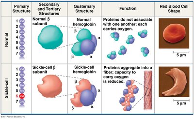
Proteins have four levels of structure, each critical for their function. Mutations can alter these structures, leading to diseases such as sickle cell anemia.
Primary Structure: Linear sequence of amino acids in a polypeptide chain.
Secondary Structure: Local folding into alpha-helices and beta-sheets, stabilized by hydrogen bonds.
Tertiary Structure: Overall 3D shape of a single polypeptide, stabilized by various interactions (hydrogen bonds, ionic bonds, hydrophobic interactions, disulfide bridges).
Quaternary Structure: Association of multiple polypeptide subunits to form a functional protein.
Sickle Cell Disease: Molecular Basis
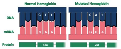
Sickle cell disease is caused by a single nucleotide mutation in the gene encoding the beta-globin subunit of hemoglobin. This mutation leads to the substitution of valine for glutamic acid, altering the protein's structure and function.
Normal Hemoglobin: Glutamic acid at position 6; hemoglobin molecules remain separate and carry oxygen efficiently.
Mutated Hemoglobin (Sickle Cell): Valine at position 6; hemoglobin molecules aggregate, distorting red blood cells into a sickle shape and impairing oxygen transport.
Membrane Structure and Function
Phospholipid Bilayer and Fluid Mosaic Model
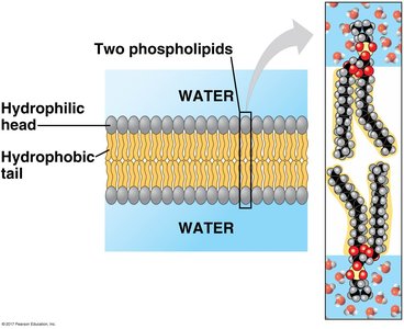
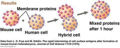
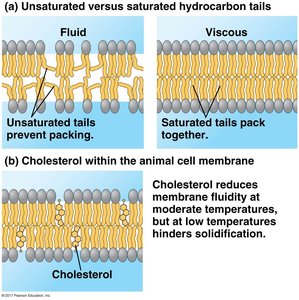
The plasma membrane is primarily composed of a phospholipid bilayer with embedded proteins, cholesterol, and carbohydrates. This structure is described by the fluid mosaic model, which emphasizes the dynamic and heterogeneous nature of the membrane.
Phospholipids: Amphipathic molecules with hydrophilic heads and hydrophobic tails, forming a bilayer in aqueous environments.
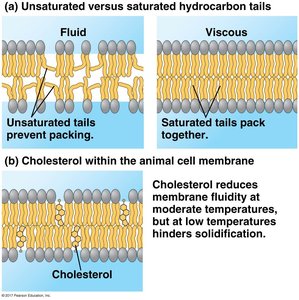
Fluidity: Membrane fluidity is influenced by temperature, fatty acid composition (saturated vs. unsaturated), and cholesterol content.
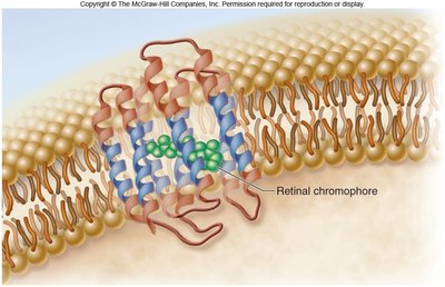
Proteins: Integral and peripheral proteins serve various functions, including transport, signaling, and cell recognition.
Membrane Proteins and Their Functions
Membrane proteins are essential for the diverse functions of biological membranes. They can act as transporters, enzymes, receptors, and more.
Transport Proteins: Facilitate movement of substances across the membrane (channels, carriers, pumps).
Enzymatic Activity: Catalyze specific reactions at the membrane surface.
Signal Transduction: Transmit signals from the extracellular environment to the cell interior.
Cell-Cell Recognition: Glycoproteins serve as identification tags for cell recognition.
Intercellular Joining: Proteins help connect adjacent cells.
Attachment: Anchor the membrane to the cytoskeleton and extracellular matrix.
Transport Across Membranes
Cells regulate the movement of substances across their membranes through various mechanisms, including passive and active transport.
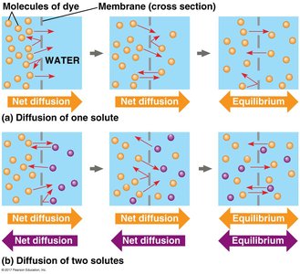
Diffusion: Movement of molecules from high to low concentration.
Facilitated Diffusion: Passive transport of molecules via protein channels or carriers.
Osmosis: Diffusion of water across a selectively permeable membrane toward higher solute concentration.
Active Transport: Movement of molecules against their concentration gradient, requiring energy (usually ATP).
Bulk Transport: Endocytosis and Exocytosis
Large molecules and particles are transported across membranes via bulk transport mechanisms.
Endocytosis: Uptake of materials into the cell by vesicle formation (includes phagocytosis, pinocytosis, and receptor-mediated endocytosis).
Exocytosis: Secretion of materials out of the cell via vesicle fusion with the plasma membrane.
Summary Table: Comparison of Animal and Plant Cells
Feature | Animal Cell | Plant Cell |
|---|---|---|
Cell Wall | No | Yes |
Chloroplasts | No | Yes |
Central Vacuole | No (small vesicles) | Yes (large) |
Lysosomes | Yes | Rare |
Shape | Round/Irregular | Rectangular/Regular |
Additional info: The notes above integrate content from the provided materials and expand with academic context to ensure completeness and clarity for college-level biology students.
