 Back
BackMembrane Structure and Function: Study Guide
Study Guide - Smart Notes
 Tailored notes based on your materials, expanded with key definitions, examples, and context.
Membrane Structure and Function
Phospholipid Bilayer and Membrane Structure
The cell membrane is primarily composed of a phospholipid bilayer, which forms the fundamental structure of all biological membranes. Phospholipids are amphipathic molecules, meaning they have both hydrophilic (water-attracting) heads and hydrophobic (water-repelling) tails. This dual nature allows them to spontaneously form bilayers in aqueous environments, with hydrophobic tails facing inward and hydrophilic heads facing outward.
Amphipathic: Molecules with both hydrophilic and hydrophobic regions.
Fluid Mosaic Model: Describes the membrane as a dynamic structure with proteins embedded in or attached to a flexible lipid bilayer.
Hydrophobic Interactions: Drive the formation of the bilayer and influence membrane properties.
Membrane fluidity is crucial for function and is influenced by temperature, fatty acid saturation, cholesterol content, and tail length:
Decreasing temperature reduces fluidity.
Unsaturated hydrocarbon chains increase fluidity due to kinks from double bonds.
Cholesterol acts as a buffer, maintaining fluidity across temperature changes.
Longer hydrocarbon tails decrease fluidity.
Membrane Proteins and Their Functions
Proteins are the 'mosaic' part of the fluid mosaic model and are essential for membrane function. They are classified as:
Integral proteins: Penetrate the hydrophobic core; often span the membrane (transmembrane proteins).
Peripheral proteins: Loosely attached to the membrane surface; involved in signaling or structural support.
Lipid-anchored proteins: Covalently attached to lipids within the bilayer.
Major functions of membrane proteins include:
Transport: Move substances across the membrane.
Enzymatic activity: Catalyze biochemical reactions.
Signal transduction: Relay signals from outside to inside the cell.
Cell-cell recognition: Identify and interact with other cells.
Intercellular joining: Connect adjacent cells.
Attachment to cytoskeleton and ECM: Maintain cell shape and stability.
Membrane Carbohydrates and Cell Recognition
Membrane carbohydrates are involved in cell-cell recognition and are found as:
Glycolipids: Carbohydrates attached to lipids.
Glycoproteins: Carbohydrates attached to proteins.
Examples include immune cell recognition and blood type determination. Glycosylation is the process of adding carbohydrate groups to proteins or lipids, which is critical for cellular identification and communication.
Selective Permeability of Membranes
Cell membranes exhibit selective permeability, allowing some substances to cross more easily than others. Small, nonpolar molecules (e.g., O2, CO2, steroids) can diffuse freely, while large or charged molecules (e.g., glucose, ions, water) require transport proteins.
Transport proteins (channels and carriers) facilitate movement of specific molecules, imparting selectivity to the membrane.
Passive and Active Transport
Transport across membranes can be passive or active:
Passive diffusion: Movement down a concentration gradient without energy or proteins.
Facilitated diffusion: Passive movement via transport proteins (channels or carriers); no energy required.
Active transport: Movement against a gradient using energy (usually ATP) and specific transport proteins (pumps).
Key differences:
Passive transport does not require energy; active transport does.
Facilitated diffusion and active transport both use proteins, but only active transport moves substances against their gradient.
Osmosis and Tonicity
Osmosis is the diffusion of water across a selectively permeable membrane. The direction of water movement depends on the relative solute concentrations (tonicity):
Isotonic: Equal solute concentration; no net water movement.
Hypertonic: Higher solute concentration outside; water leaves the cell.
Hypotonic: Lower solute concentration outside; water enters the cell.
Consequences for cells:
Animal cells: Crenation (shrink) in hypertonic, lysis (burst) in hypotonic, normal in isotonic.
Plant cells: Plasmolysis in hypertonic, turgor in hypotonic, flaccid in isotonic.
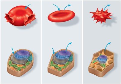
Image explanation: The image shows animal and plant cells in isotonic, hypotonic, and hypertonic solutions. Arrows indicate the direction of water movement. Animal cells undergo crenation (shrink) in hypertonic, lysis (burst) in hypotonic, and remain normal in isotonic. Plant cells experience plasmolysis in hypertonic, turgor in hypotonic, and are flaccid in isotonic solutions.
Facilitated Diffusion: Channels vs. Carriers
Facilitated diffusion is a type of passive transport that uses proteins to move substances across the membrane. There are two main types of transport proteins:
Channel proteins: Provide open passageways for specific molecules or ions.
Carrier proteins: Bind to molecules and change shape to shuttle them across the membrane; specificity is determined by the shape of the binding site.
Active Transport: Primary and Secondary
Active transport moves substances against their concentration gradients using energy. There are two main types:
Primary active transport: Directly uses ATP to transport molecules (e.g., sodium-potassium pump).
Secondary active transport (cotransport): Uses the energy from the gradient created by primary active transport to move other substances.
The sodium-potassium pump is a classic example, moving 3 Na+ out and 2 K+ into the cell using ATP. Cotransporters move two substances simultaneously, one down its gradient and one against.
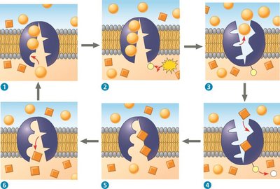
Image explanation: The image illustrates the steps of the sodium-potassium pump: binding of Na+, phosphorylation by ATP, conformational change, release of Na+, binding of K+, dephosphorylation, and release of K+ into the cytoplasm. This cycle maintains essential ion gradients across the membrane.
Bulk Transport: Exocytosis and Endocytosis
Large particles and macromolecules cross membranes via bulk transport, which requires energy (active transport):
Exocytosis: Vesicles fuse with the membrane to release contents outside the cell (e.g., neurotransmitter release).
Endocytosis: Membrane engulfs material to bring it into the cell. Types include:
Phagocytosis: Engulfment of solid particles (e.g., white blood cells ingesting bacteria).
Pinocytosis: Uptake of extracellular fluid.
Receptor-mediated endocytosis: Specific uptake via receptor proteins (e.g., cholesterol uptake).
All bulk transport processes are active, requiring energy input.
Key Terms and Definitions
Equilibrium: State where concentrations are equal across a membrane.
Concentration gradient: Difference in concentration across a space or membrane.
Uniporter: Transports one type of molecule.
Symporter: Transports two molecules in the same direction.
Antiporter: Transports two molecules in opposite directions.
Summary Table: Types of Membrane Transport
Transport Type | Energy Required? | Protein Required? | Direction Relative to Gradient | Example |
|---|---|---|---|---|
Passive Diffusion | No | No | Down | O2, CO2 |
Facilitated Diffusion | No | Yes | Down | Glucose, H2O (aquaporins) |
Active Transport | Yes | Yes | Against | Na+/K+ pump |
Bulk Transport | Yes | Yes (vesicles) | Varies | Exocytosis, Endocytosis |
Key Equations
Osmosis (Water Potential):
Where is water potential, is solute potential, and is pressure potential.
Sodium-Potassium Pump Stoichiometry:
Additional info: This guide expands on the provided study guide with academic context, definitions, and examples to ensure a comprehensive understanding of membrane structure and function for college-level biology students.
