 Back
BackMembrane Structure and Function: Study Notes
Study Guide - Smart Notes
 Tailored notes based on your materials, expanded with key definitions, examples, and context.
Membrane Structure and Function
Biological Membranes: Structure and Composition
Biological membranes are essential structures that define cell boundaries and regulate the movement of substances into and out of cells. The primary component of these membranes is the phospholipid bilayer, which is interspersed with proteins, cholesterol, and carbohydrates, forming a dynamic and functional barrier.
Phospholipids are amphipathic molecules, meaning they have both hydrophilic (water-attracting) and hydrophobic (water-repelling) regions.
The fluid mosaic model describes the membrane as a fluid structure with a "mosaic" of various proteins embedded in or attached to a double layer (bilayer) of phospholipids.
Cholesterol is interspersed within the bilayer, modulating membrane fluidity and stability.
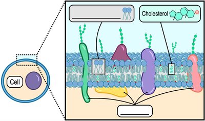
Example: The major components of biological membranes are phospholipids, proteins, and cholesterol.
Types of Membrane Proteins
Membrane proteins are crucial for the diverse functions of biological membranes. They are classified based on their association with the lipid bilayer:
Integral membrane proteins are embedded within the lipid bilayer and often span the entire membrane (transmembrane proteins).
Peripheral membrane proteins are attached to the exterior or interior surfaces of the membrane and do not penetrate the hydrophobic core.
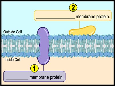
Example: Integral proteins span the membrane, while peripheral proteins are attached to the membrane's surface.
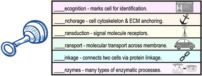
Functions of Membrane Proteins
Membrane-associated proteins perform a wide variety of functions, including:
Recognition: Marking cells for identification (e.g., immune response).
Anchorage: Attaching the cytoskeleton to the extracellular matrix (ECM).
Signal transduction: Acting as receptors for signal molecules.
Transport: Facilitating the movement of substances across the membrane.
Linkage: Connecting adjacent cells via protein linkages.
Enzymatic activity: Catalyzing specific reactions at the membrane surface.
Membrane Transport Mechanisms
Concentration Gradients and Diffusion
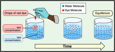
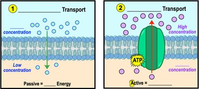
A concentration gradient exists when there is a difference in the concentration of a substance across a space or a membrane. Molecules naturally move from areas of high concentration to areas of low concentration (down their gradient), a process called diffusion.
Movement down the gradient does not require energy.
Movement against the gradient requires energy input.
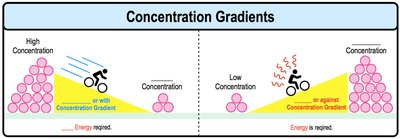
Example: Diffusion of dye in water demonstrates molecules moving from high to low concentration until equilibrium is reached.
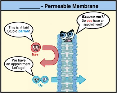
Selective Permeability of Membranes
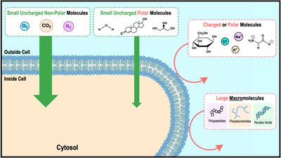
Biological membranes are selectively permeable, meaning they allow some substances to cross more easily than others. Small, nonpolar molecules (e.g., O2, CO2) can diffuse freely, while large or charged molecules require assistance.
CAN Freely Diffuse | Cannot Freely Diffuse |
|---|---|
Small | LARGE |
Uncharged | (+/-) Charged |
Nonpolar/Hydrophobic | Polar/Hydrophilic |

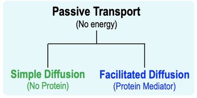
Types of Membrane Transport
Transport across membranes can be divided into passive (no energy required) and active (energy required) processes. Bulk transport mechanisms include endocytosis and exocytosis for large molecules.
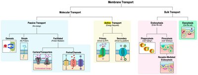
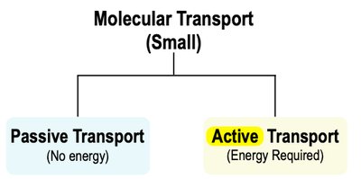
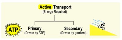
Passive vs. Active Transport
Passive transport moves substances down their concentration gradient without energy input. Active transport moves substances against their gradient, requiring energy (usually from ATP).
Passive: High to low concentration, no energy.
Active: Low to high concentration, requires ATP.
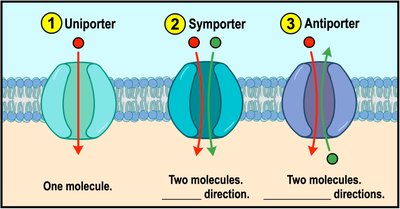
Classes of Membrane Transport Proteins
Transport proteins facilitate the movement of molecules across membranes and are classified as:
Uniporters: Transport one molecule at a time in one direction.
Symporters: Cotransport two or more molecules in the same direction.
Antiporters: Cotransport two or more molecules in opposite directions.
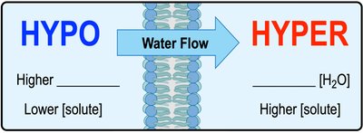
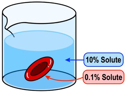
Osmosis and Tonicity
Osmosis
Osmosis is the passive diffusion of water across a semi-permeable membrane. The direction of water movement depends on the relative concentration of solutes (tonicity) on either side of the membrane.
Hypotonic: Lower solute concentration outside the cell; water enters the cell.
Isotonic: Equal solute concentration; no net water movement.
Hypertonic: Higher solute concentration outside the cell; water leaves the cell.

Simple and Facilitated Diffusion
Simple Diffusion
Simple diffusion is the direct movement of small, uncharged molecules (e.g., O2, CO2) through the lipid bilayer without the need for transport proteins or energy.
Facilitated Diffusion
Facilitated diffusion is the passive movement of charged or polar molecules across the membrane via specific transport proteins (channels or carriers). No energy is required, but a protein mediator is necessary.

Active Transport
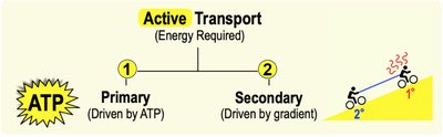
Primary and Secondary Active Transport
Active transport moves molecules against their concentration gradients and requires energy. There are two main types:
Primary active transport: Directly uses ATP to transport molecules (e.g., Na+/K+ pump).
Secondary active transport: Uses the energy stored in concentration gradients established by primary active transport to move other substances.
Example: Sodium-Potassium Pump
The Na+/K+ pump is a classic example of primary active transport, exporting three sodium ions out of the cell and importing two potassium ions into the cell, using energy from ATP hydrolysis. This process maintains essential electrochemical gradients across the plasma membrane.
Bulk Transport: Endocytosis and Exocytosis
Endocytosis
Endocytosis is the process by which cells engulf large particles or macromolecules by wrapping the plasma membrane around them and forming a vesicle. Types include:
Phagocytosis: "Cell eating" – uptake of large particles or cells.
Pinocytosis: "Cell drinking" – uptake of extracellular fluid and dissolved solutes.
Receptor-mediated endocytosis: Specific uptake of molecules via receptor proteins.
Exocytosis
Exocytosis is the process by which cells expel materials in vesicles that fuse with the plasma membrane, releasing their contents outside the cell. This is important for secretion of hormones, neurotransmitters, and waste products.
Additional info: These processes are essential for nutrient uptake, immune responses, and intercellular communication.
