 Back
BackMembrane Structure, Function, and Cell Communication: Study Notes
Study Guide - Smart Notes
 Tailored notes based on your materials, expanded with key definitions, examples, and context.
Membrane Structure and Function
Chemical Composition of Cellular Membranes
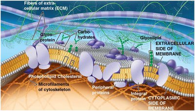
The plasma membrane is a dynamic structure that separates the cell from its environment and regulates the movement of substances in and out of the cell. It is primarily composed of lipids, proteins, and carbohydrates.
Phospholipids are the main lipid component, forming a bilayer with hydrophilic heads facing outward and hydrophobic tails inward.
Proteins are embedded within or attached to the bilayer, serving various functions such as transport, signaling, and structural support.
Carbohydrates are attached to lipids (glycolipids) or proteins (glycoproteins) and play roles in cell recognition and signaling.
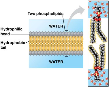
Amphipathic molecules have both hydrophilic and hydrophobic regions, a property crucial for membrane structure.
The Fluid Mosaic Model
The fluid mosaic model describes the plasma membrane as a mosaic of proteins floating in or on the fluid lipid bilayer. This model explains the flexibility and dynamic nature of membranes.
Proteins are not randomly distributed; they often form functional groups.
Membrane fluidity is maintained by weak hydrophobic interactions among lipids and proteins.
Lipids and some proteins can move laterally within the membrane; flip-flop between layers is rare.
Factors Affecting Membrane Fluidity
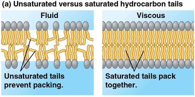
Membrane fluidity is essential for proper function and is influenced by lipid composition and temperature.
Unsaturated fatty acid tails prevent tight packing, increasing fluidity.
Saturated fatty acid tails pack closely, making the membrane more viscous.
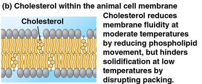
Cholesterol acts as a fluidity buffer in animal cells, restraining movement at high temperatures and preventing solidification at low temperatures.
Membrane Proteins and Their Functions
Membrane proteins determine most of the membrane's specific functions. They can be classified as:
Peripheral proteins: Loosely bound to the membrane surface.
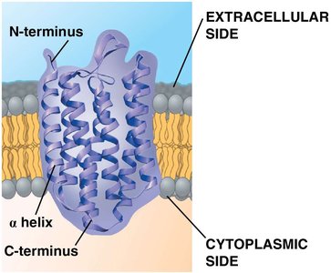
Integral proteins: Penetrate the hydrophobic core; transmembrane proteins span the entire membrane.
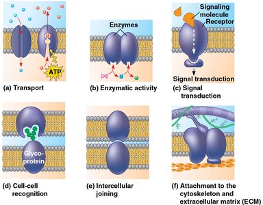
Functions include transport, enzymatic activity, signal transduction, cell-cell recognition, intercellular joining, and attachment to the cytoskeleton and extracellular matrix (ECM).
Membrane Carbohydrates and Cell-Cell Recognition
Carbohydrates on the cell surface function as markers for cell recognition. They are usually short, branched chains attached to lipids (glycolipids) or proteins (glycoproteins).
These markers are important for immune response and tissue organization.
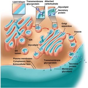
Sidedness of Membranes
Membranes have distinct inside and outside faces, with asymmetrical distribution of proteins, lipids, and carbohydrates. This sidedness is established during membrane synthesis in the endoplasmic reticulum and Golgi apparatus.
Selective Permeability of Membranes
The plasma membrane is selectively permeable, allowing some substances to cross more easily than others.
Hydrophobic (nonpolar) molecules (e.g., O2, CO2) pass through rapidly.
Hydrophilic (polar) molecules (e.g., ions, sugars) require transport proteins to cross efficiently.
Transport Across Membranes
Substances cross membranes by several mechanisms:
Simple diffusion: Movement down a concentration gradient without energy input.
Facilitated diffusion: Passive transport aided by transport proteins (channel or carrier proteins).
Active transport: Movement against a concentration gradient, requiring energy (usually ATP) and carrier proteins.
Bulk transport: Movement of large molecules via vesicles (endocytosis and exocytosis).

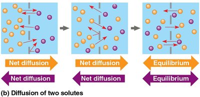
Passive Transport: Diffusion and Osmosis
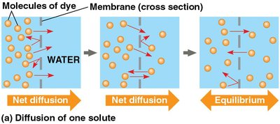
Passive transport does not require energy. Diffusion is the movement of molecules from high to low concentration. Osmosis is the diffusion of water across a selectively permeable membrane.
Water moves from areas of low solute concentration to high solute concentration until equilibrium is reached.
Tonicity and Water Balance
Tonicity describes a solution's ability to cause a cell to gain or lose water:
Isotonic: Solute concentration is equal inside and outside; no net water movement.
Hypertonic: Higher solute concentration outside; cell loses water and shrivels.
Hypotonic: Lower solute concentration outside; cell gains water and may burst.
Cells without walls (e.g., animal cells) are sensitive to tonicity, while cells with walls (e.g., plant cells) are protected by turgor pressure.
Facilitated Diffusion
Facilitated diffusion uses transport proteins to move substances down their concentration gradients without energy input.
Channel proteins (e.g., aquaporins, ion channels) provide corridors for specific molecules or ions.
Carrier proteins change shape to shuttle molecules across the membrane.
Active Transport
Active transport moves substances against their concentration gradients using energy, typically from ATP hydrolysis.
Examples include the sodium-potassium pump in animal cells and the proton pump in plants, fungi, and bacteria.
Active transport maintains essential differences in ion concentrations across membranes.
Bulk Transport: Endocytosis and Exocytosis
Large molecules cross the membrane via vesicles in processes called endocytosis (import) and exocytosis (export).
Exocytosis: Vesicles fuse with the plasma membrane to release contents outside the cell.
Endocytosis: The cell takes in macromolecules by forming vesicles from the plasma membrane. Types include phagocytosis, pinocytosis, and receptor-mediated endocytosis.
Cell Communication
Overview of Cell Signaling
Cells communicate through signaling molecules that trigger responses in target cells. This process is essential for coordination in multicellular organisms and is conserved across evolution.
Signaling can be local (paracrine, synaptic) or long-distance (hormonal).
Quorum sensing in bacteria is an example of cell signaling in prokaryotes.
The Three Stages of Cell Signaling
Cell signaling involves three main steps:
Reception: The target cell detects a signaling molecule (ligand) that binds to a receptor protein.
Transduction: The receptor changes shape, initiating a signal transduction pathway, often involving multiple steps.
Response: The transduced signal triggers a specific cellular response.
Types of Receptors
Cell surface receptors: Bind water-soluble ligands; include G protein-coupled receptors (GPCRs), receptor tyrosine kinases (RTKs), and ion channel receptors.
Intracellular receptors: Located in the cytoplasm or nucleus; bind small or hydrophobic ligands (e.g., steroid hormones).
Ligand type determines receptor location: hydrophilic ligands bind cell surface receptors, while hydrophobic ligands bind intracellular receptors.
Apoptosis: Programmed Cell Death
Apoptosis is a form of programmed cell death essential for development and maintenance in multicellular organisms.
Triggered by internal or external signals, leading to activation of caspases (proteases) that dismantle the cell.
Prevents damage to neighboring cells by containing cellular contents.
Examples: Formation of fingers and toes in humans, removal of damaged or infected cells, and prevention of cancer.
Additional info: Apoptosis is implicated in diseases such as Parkinson's and Alzheimer's when dysregulated.
