 Back
Membrane Transport and Cell Signaling – Study Notes (Campbell Biology in Focus, Ch. 5)
Study Guide - Smart Notes
 Tailored notes based on your materials, expanded with key definitions, examples, and context.
Membrane Transport and Cell Signaling
Introduction to Membrane Function
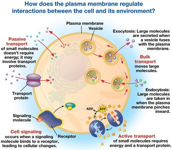
The plasma membrane is a fundamental cellular structure that regulates the movement of substances into and out of the cell, maintaining homeostasis and enabling communication with the environment. It is composed primarily of a phospholipid bilayer with embedded proteins, carbohydrates, and cholesterol, forming a dynamic and selectively permeable barrier.
Structure of Cellular Membranes
Fluid Mosaic Model
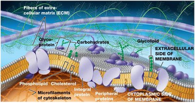
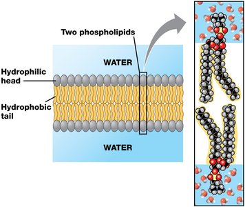
The fluid mosaic model describes the plasma membrane as a mosaic of protein molecules bobbing in a fluid bilayer of phospholipids. Phospholipids are amphipathic, containing both hydrophilic (water-attracting) heads and hydrophobic (water-repelling) tails, which allows them to form stable bilayers in aqueous environments.
Phospholipid bilayer: Forms the basic structure of the membrane, with hydrophobic tails facing inward and hydrophilic heads facing outward.
Membrane proteins: Integral and peripheral proteins are interspersed throughout the bilayer, contributing to membrane function.
Carbohydrates: Attached to proteins (glycoproteins) or lipids (glycolipids) on the extracellular surface, important for cell recognition.
Cholesterol: Modulates membrane fluidity and stability.
Membrane Fluidity
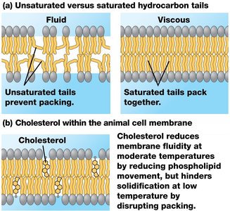
Membrane fluidity is essential for proper function, allowing for the movement of proteins and lipids within the bilayer. Fluidity is influenced by temperature, the composition of fatty acid tails, and cholesterol content.
Unsaturated hydrocarbon tails: Increase fluidity by preventing tight packing.
Saturated hydrocarbon tails: Decrease fluidity by allowing tighter packing.
Cholesterol: Reduces fluidity at moderate temperatures but prevents solidification at low temperatures.
Membrane Proteins
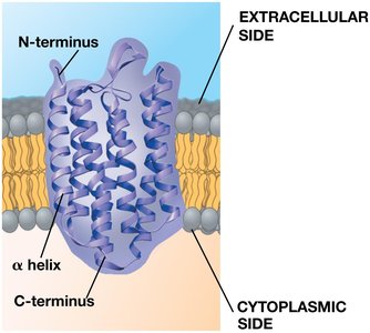
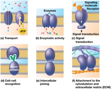
Proteins embedded in the membrane perform a variety of functions, including transport, enzymatic activity, signal transduction, cell-cell recognition, intercellular joining, and attachment to the cytoskeleton and extracellular matrix (ECM).
Integral proteins: Penetrate the hydrophobic core; many are transmembrane proteins.
Peripheral proteins: Loosely bound to the membrane surface.
Membrane Carbohydrates and Cell Recognition
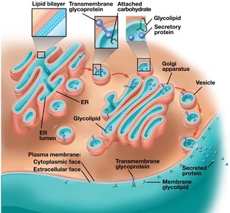
Carbohydrates on the extracellular surface of the plasma membrane are crucial for cell-cell recognition. They are covalently bonded to lipids (glycolipids) or proteins (glycoproteins) and vary among species, individuals, and cell types.
Synthesis and Sidedness of Membranes
Membranes have distinct inside and outside faces, established during their synthesis in the endoplasmic reticulum (ER) and Golgi apparatus. This asymmetry is critical for membrane function.
Membrane Permeability and Transport
Selective Permeability
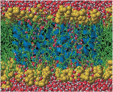
The plasma membrane is selectively permeable, allowing some substances to cross more easily than others. Hydrophobic (nonpolar) I molecules pass through easily, while hydrophilic (polar) molecules and ions require transport proteins.
Transport Proteins
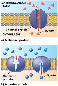
Transport proteins facilitate the movement of hydrophilic substances across the membrane. Channel proteins provide hydrophilic tunnels, while carrier proteins bind and shuttle molecules across.
Channel proteins: Form pores for specific molecules or ions (e.g., aquaporins for water). |
Carrier proteins: Undergo shape changes to move substances across the membrane.
Passive Transport
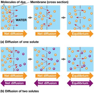
Diffusion
Diffusion is the movement of molecules from an area of higher concentration to an area of lower concentration, driven by the concentration gradient. It is a form of passive transport, requiring no energy input.
At dynamic equilibrium, molecules continue to move, but there is no net change in concentration.
Osmosis
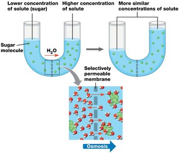
Osmosis is the diffusion of free water across a selectively permeable membrane. Water moves from regions of lower solute concentration to regions of higher solute concentration until equilibrium is reached.
Tonicity and Water Balance
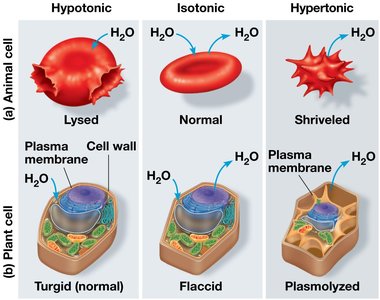
Tonicity describes the ability of a solution to cause a cell to gain or lose water:
Isotonic: No net water movement; cell remains stable.
Hypertonic: Cell loses water; shrivels.
Hypotonic: Cell gains water; may lyse (burst).
Osmoregulation
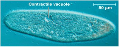
Cells without walls must regulate water balance to survive in hypotonic or hypertonic environments. Some protists, such as Paramecium caudatum, use contractile vacuoles to expel excess water.
Facilitated Diffusion
Facilitated diffusion is a type of passive transport aided by proteins. Channel proteins and carrier proteins help specific molecules cross the membrane without energy input.
Active Transport
Mechanism and Importance
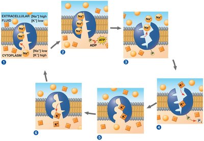
Active transport moves substances against their concentration gradients, requiring energy (usually from ATP). This process allows cells to maintain internal concentrations of small molecules that differ from those in the environment.
Sodium-potassium pump: Exchanges Na+ for K+ across the plasma membrane of animal cells, maintaining electrochemical gradients essential for cell function.
Summary Table: Types of Membrane Transport
Type | Energy Required? | Direction | Example |
|---|---|---|---|
Simple Diffusion | No | Down gradient | O2, CO2 |
Facilitated Diffusion | No | Down gradient | Glucose, ions via channels |
Active Transport | Yes (ATP) | Against gradient | Na+/K+ pump |
Bulk Transport
Exocytosis and Endocytosis
Large molecules, such as proteins and polysaccharides, cross the membrane via bulk transport mechanisms that require energy.
Exocytosis: Vesicles fuse with the plasma membrane to release contents outside the cell.
Endocytosis: The cell takes in molecules by forming vesicles from the plasma membrane. Types include phagocytosis, pinocytosis, and receptor-mediated endocytosis.
Cell Signaling
Overview of Cell Communication
Cells communicate through signaling pathways that involve the plasma membrane. Communication can be local (direct contact or local regulators) or long-distance (hormones).
Reception: Detection of a signaling molecule by a receptor.
Transduction: Conversion of the signal to a cellular response via a signal transduction pathway.
Response: Cellular activity in response to the signal.
Types of Receptors
G protein-coupled receptors (GPCRs): Work with G proteins to transmit signals.
Ligand-gated ion channels: Open or close in response to binding of a signaling molecule, allowing ions to pass through.
Intracellular receptors: Located in the cytoplasm or nucleus; activated by small or hydrophobic molecules (e.g., steroid hormones).
Signal Transduction Pathways
Signal transduction often involves multiple steps, including protein phosphorylation cascades and the use of second messengers such as cyclic AMP (cAMP) and calcium ions (Ca2+).
Protein kinases: Enzymes that transfer phosphate groups to proteins (phosphorylation).
Protein phosphatases: Remove phosphate groups (dephosphorylation).
Second messengers: Small molecules that relay signals inside the cell (e.g., cAMP, Ca2+).
Cellular Responses
The final outcome of cell signaling can be regulation of gene expression (transcription) or changes in cytoplasmic activities, such as enzyme activity or ion channel opening.
