 Back
BackMicrobial Life: Prokaryotes and Protists – Structured Study Notes
Study Guide - Smart Notes
 Tailored notes based on your materials, expanded with key definitions, examples, and context.
Microbial Life: Prokaryotes and Protists
Introduction to Prokaryotes
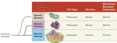
Prokaryotes are unicellular organisms that lack a membrane-bound nucleus and organelles. They are classified into two domains: Bacteria and Archaea. Prokaryotes are the most abundant and diverse organisms on Earth, playing essential roles in ecological and evolutionary processes.
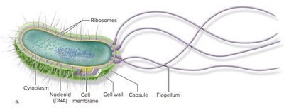
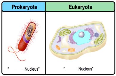
Prokaryotic Cells: Do not have a nucleus; DNA is found in a region called the nucleoid.
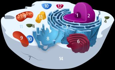
Eukaryotic Cells: Have a nucleus and other membrane-bound organelles.
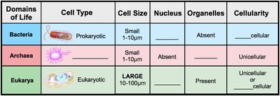
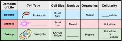
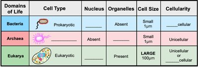
Domains of Life: Bacteria and Archaea are prokaryotic; Eukarya are eukaryotic.
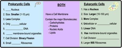
Prokaryotic vs. Eukaryotic Cells
Prokaryotic and eukaryotic cells differ in several fundamental ways, including cell size, complexity, presence of nucleus, and organelles.
Size: Prokaryotes are smaller (1-10 μm); eukaryotes are larger (10-100 μm).
Nucleus: Prokaryotes lack a nucleus; eukaryotes possess a nucleus.
Organelles: Prokaryotes lack membrane-bound organelles; eukaryotes have them.
Ribosomes: Prokaryotes have smaller 70S ribosomes; eukaryotes have larger 80S ribosomes.
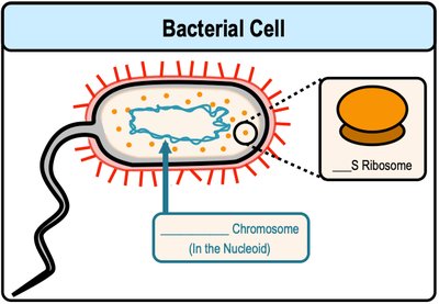
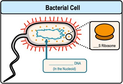
Features of Bacterial Cells
Bacteria are characterized by their unique cellular structures and functions.
DNA: Circular DNA located in the nucleoid.
Cell Division: Binary fission.
Ribosomes: Small 70S ribosomes.
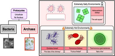
Introduction to Archaea
Archaea are prokaryotic organisms with unique features distinguishing them from bacteria.
Cell Structure: Prokaryotic, but with unique ribosomal RNA sequences.
Cell Wall: Lacks peptidoglycan.
Extremophiles: Many archaea thrive in extreme environments (e.g., high temperature, salinity).
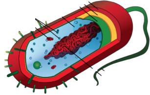
Prokaryotic Cell Structure
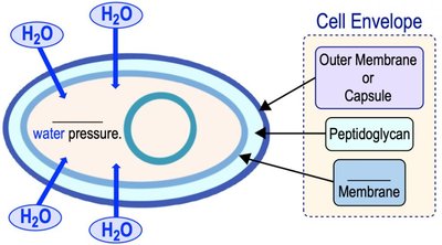
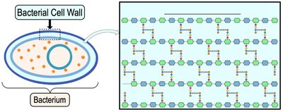
Bacterial Cell Walls and Envelope
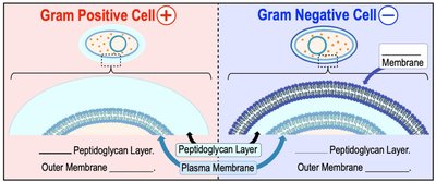
Bacterial cell walls provide structural support and protection against osmotic pressure. The cell envelope includes the cell wall, cell membrane, and sometimes an outer membrane.
Cell Wall: Semi-rigid layer outside the cell membrane.
Peptidoglycan: Main component of bacterial cell walls; provides rigidity.
Cell Envelope: Includes cell wall, membrane, and outer membrane (if present).
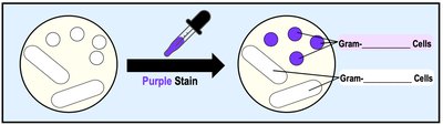
Gram-Positive vs. Gram-Negative Bacteria
Bacteria are classified based on their cell wall structure and response to Gram staining.
Gram-Positive: Thick peptidoglycan layer; retains purple stain.
Gram-Negative: Thin peptidoglycan layer and complex outer membrane; does not retain stain.
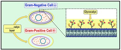
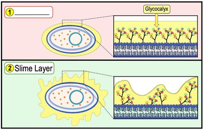
The Glycocalyx: Capsules and Slime Layers
The glycocalyx is a sticky, gel-like layer surrounding bacterial cells, aiding in attachment and protection.
Capsules: Highly organized, dense polysaccharide layer tightly anchored to the cell.
Slime Layers: Unorganized, easily removable polysaccharide layer.
Functions: Protection from dehydration, attachment to surfaces, and nutrient collection.
Pili and Fimbriae
Pili and fimbriae are filamentous protein structures on the surface of bacterial cells, involved in adhesion and motility.
Pili: Longer, fewer per cell; involved in motility and DNA transfer (conjugation).
Fimbriae: Shorter, more numerous; involved in biofilm formation and adhesion.
Endospores
Endospores are dormant, highly resistant cells produced by some bacteria (e.g., Bacillus, Clostridium) under adverse conditions.
Resistant to: Heat, chemicals, nutrient depletion.
Function: Survival, not reproduction.
Prokaryotic Motility
Chemotaxis and Flagella
Motile prokaryotes move toward favorable environments (chemoattractants) or away from harmful substances (chemorepellents) using flagella.
Chemotaxis: Movement in response to chemical gradients.
Flagella: Long, filamentous proteins driving motility; composed of filament, hook, and basal body.
Prokaryotic Reproduction
Plasmids
Plasmids are small, circular, double-stranded DNA molecules that replicate independently of the bacterial chromosome.
Contain: Genes for antibiotic resistance, conjugation, and other functions.
Plasmid Curing: Loss of plasmid from the cell.
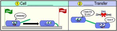
Binary Fission
Binary fission is the primary method of prokaryotic asexual reproduction, resulting in two genetically identical daughter cells.
Process: DNA replication, cell elongation, division.
Horizontal Gene Transfer
Horizontal gene transfer allows prokaryotes to acquire new traits and genetic diversity through three main mechanisms:
Transformation: Uptake of free DNA from the environment by competent cells.
Transduction: DNA transfer mediated by bacteriophage viruses.
Conjugation: Direct DNA transfer between cells via cell-to-cell contact, often involving a sex pilus.
Prokaryotic Metabolism
Nutritional Factors and Diversity
Microbes are classified based on their energy, electron, and carbon sources:
Energy Source: Phototrophs (sunlight), Chemotrophs (chemical compounds).
Electron Source: Lithotrophs (inorganic molecules), Organotrophs (organic molecules).
Carbon Source: Autotrophs (CO2), Heterotrophs (organic molecules).
Oxygen Requirements
Microbes are classified by their oxygen requirements:
Aerobes: Require O2.
Anaerobes: Grow without O2.
Facultative: Can grow with or without O2.
Biofilms
Biofilms are complex communities of microbes encased in a polysaccharide matrix, adhering to surfaces and often causing illness.
Extracellular Polymeric Substances (EPS): Matrix of polysaccharides, proteins, glycoproteins, glycolipids, and lipids.
Prokaryotic Diversity
Major Prokaryotic Lineages
Prokaryotes are classified into several major lineages:
Proteobacteria: Diverse, gram-negative, includes nitrogen-fixing and pathogenic species.
Chlamydiae: Gram-negative, lack peptidoglycan, obligate intracellular parasites.
Spirochetes: Gram-negative, corkscrew-shaped, many pathogenic.
Cyanobacteria: Gram-negative, photoautotrophs, oxygenic photosynthesis.
Actinobacteria: High-GC gram-positive, includes antibiotic-producing Streptomyces.
Firmicutes: Low-GC gram-positive, includes Lactobacillus.
Environmental Factors
Microbes are classified by their tolerance to environmental factors such as salt, temperature, and pH.
Halotolerants: Tolerate moderate salt.
Halophiles: Require high salt.
Extreme Halophiles: Require very high salt.
Thermophiles: Thrive in high temperatures.
Acidophiles: Thrive in acidic environments.
Prokaryotes in the Environment
Ecological Roles
Bacteria and archaea drive nutrient and energy cycles, including nitrogen fixation and oxygen production. They are also crucial in the internal environments of humans and animals.
The Human Microbiome
The human microbiome consists of communities of microbes living in symbiotic relationships with humans, classified as resident or transient microbiota.
Resident Microbiota: Persist long-term.
Transient Microbiota: Temporary, often pathogenic.
Pathogenic Toxins and Virulence
Pathogens produce toxins (exotoxins and endotoxins) that damage host tissues. Virulence factors are traits that promote pathogenicity.
Exotoxins: Soluble proteins released by pathogens.
Endotoxins: Lipopolysaccharide (LPS) in gram-negative bacteria.
Virulence Factors: Traits such as adhesins, capsules, toxins, and proteases.
Introduction to Protists
What is a Protist?
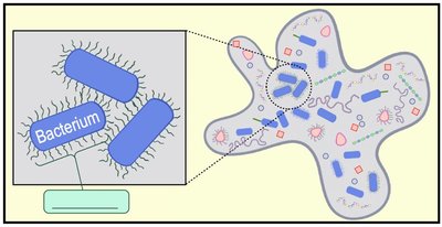
Protists are a diverse group of eukaryotic organisms that are not plants, animals, or fungi. Most eukaryotes are protists, and they exhibit a wide range of structures and functions.
Features: Membrane-bound nucleus, standard eukaryotic organelles, mostly unicellular, aquatic or moist environments.
Nutrition: Heterotrophic, photosynthetic, or mixotrophic.
Evolution of Protists
Endosymbiosis played a key role in the evolution of eukaryotes and protists.
Primary Endosymbiosis: Host cell engulfs a prokaryotic cell.
Secondary Endosymbiosis: Host cell engulfs a eukaryotic cell, leading to organelles with multiple membranes.
Protist Life Cycles
Protists exhibit diverse life cycles, including sexual and asexual reproduction, alternation of generations, and multiple hosts.
Plasmodium (Malaria): Requires mosquito and human hosts; both sexual and asexual stages.
Laminaria: Alternation of generations between diploid sporophyte and haploid gametophyte.
Paramecium: Conjugation (sexual exchange) and binary fission (asexual reproduction).
Eukaryotic Supergroups
Protists are classified into four supergroups based on genetic and morphological evidence:
Excavata
SAR
Archaeplastida
Unikonta
Supergroups emphasize evolutionary relationships and accommodate updates from new phylogenetic discoveries.
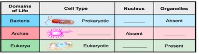
Domain | Cell Type | Nucleus | Organelles |
|---|---|---|---|
Bacteria | Prokaryotic | Absent | Absent |
Archaea | Prokaryotic | Absent | Absent |
Eukarya | Eukaryotic | Present | Present |
Cell Type | Size | Nucleus | Organelles | Cellularity |
|---|---|---|---|---|
Prokaryotic | Small (1-10 μm) | Absent | Absent | Unicellular |
Eukaryotic | Large (10-100 μm) | Present | Present | Unicellular or multicellular |
Gram Type | Peptidoglycan Layer | Outer Membrane |
|---|---|---|
Gram Positive | Thick | Absent |
Gram Negative | Thin | Present |
*Additional info: Academic context and explanations have been expanded for completeness and clarity. Tables have been recreated and logically inferred where necessary. Only images directly relevant to the explanation have been included.*
