 Back
Microscopy and Diversity of Life: Study Notes for General Biology
Study Guide - Smart Notes
 Tailored notes based on your materials, expanded with key definitions, examples, and context.
Microscopy and Diversity of Life
Introduction to Scientific Inquiry and Microscopy
Scientific inquiry in biology involves both controlled experiments and careful observation of living organisms. Microscopy is a fundamental tool that allows biologists to observe cells and microorganisms, enhancing our understanding of the diversity of life. Proper use of the compound light microscope and the development of scientific observation skills are essential for studying biological specimens.
Microscopy
Setting Up and Stowing the Compound Light Microscope
Preparation: Remove the dust cover, check for damage, and clean the stage and lenses with appropriate materials.
Operation: Turn on the light source and follow the instructor’s directions for use.
After Use: Turn off the light, remove slides, clean lenses (especially after oil immersion), set the scanning lens in place, lower and center the stage, and store the power cord properly.
Parts of the Compound Light Microscope
Understanding the function of each part of the microscope is crucial for effective use and accurate observation.
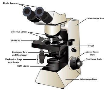
Ocular Lens: The eyepiece through which you view the specimen; typically provides 10× magnification.
Objective Lenses: Multiple lenses (e.g., 4×, 10×, 40×, 100×) that provide varying levels of magnification.
Stage: Platform where the slide is placed for observation.
Coarse and Fine Focus Knobs: Used to bring the specimen into general and sharp focus, respectively.
Light Source: Illuminates the specimen from below.
Diaphragm and Condenser: Control the amount and focus of light reaching the specimen.
Mechanical Stage: Allows precise movement of the slide.
Base and Arm: Provide support and structure to the microscope.
Microscopy Concepts and Principles
Magnification: Enlargement of the specimen; total magnification is the product of the ocular and objective lens magnifications. Equation:
Resolving Power: Ability to distinguish two close objects as separate; improved with immersion oil at high magnification.
Contrast: Difference between the specimen and background; enhanced by staining.
Depth of Field: Thickness of the specimen in focus at one time; decreases as magnification increases.
Field of View: Area visible through the microscope; decreases with higher magnification.
Working Distance: Distance between the objective lens and the slide; decreases as magnification increases.
Example: As you increase magnification from 4× to 100×, the field of view, resolution, and depth of field all decrease, allowing you to see finer details but a smaller area and thinner section of the specimen.
Cellular Diversity
Introduction to Cells
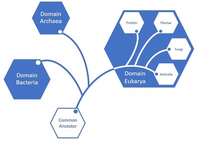
All living things are composed of cells, the basic unit of life. Cells share four essential features: a cell membrane, cytosol, DNA, and ribosomes. These features reflect the unity of life and support the theory of evolution from a common ancestor.
Prokaryotic Cells: Lack membrane-bound organelles; DNA is in a nucleoid region. Found in Domains Archaea and Bacteria.
Eukaryotic Cells: Have membrane-bound organelles, including a nucleus containing DNA. Found in Domain Eukarya (Kingdoms Plantae, Fungi, Animalia, and Protists).
Survey of Eukaryotes
Protists
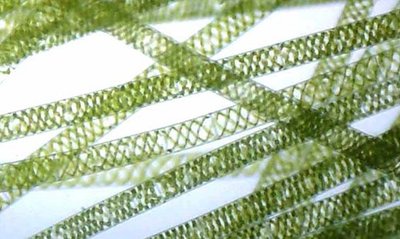
Protists are mostly unicellular eukaryotes, though some form colonies or are multicellular. Some, like Spirogyra crassa, are photosynthetic and have distinctive spiral chloroplasts.
Observation: Use a compound microscope to view prepared slides of Spirogyra crassa and focus on the spiral chloroplasts.
Microscopy Skills: As you increase magnification, the field of view and depth of field decrease, but resolution increases, allowing you to see more detail in the chloroplasts.
Fungi
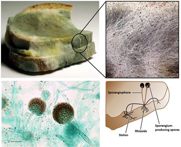
Fungi are multicellular eukaryotic heterotrophs that absorb nutrients. Rhizopus stolonifer (black bread mold) is a common example, with hyphae that penetrate substrates and sporangia that produce spores.
Observation: View prepared slides of Rhizopus stolonifer to identify hyphae and sporangia.
Staining: Stains are used to enhance visibility of fungal structures under the microscope.
Plants
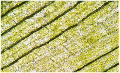
Plants are multicellular, photosynthetic eukaryotes. Elodea is a common aquatic plant used to study plant cell structure, especially chloroplasts, which are visible as green organelles inside the cells.
Wet Mount Preparation: Place a leaf in water on a slide, add a coverslip, and use iodine stain to enhance visibility.
Observation: Use the 40× objective to observe and label the cell wall and chloroplasts.
Animals
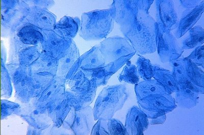
Animals are multicellular eukaryotes. Animal cells lack cell walls and chloroplasts. Human cheek cells are a common specimen for observing animal cell structure, including the nucleus and cytoplasm.
Wet Mount Preparation: Scrape the inside of the cheek, mix with water on a slide, add a coverslip, and stain with methylene blue.
Observation: Use the microscope to identify the nucleus and other visible organelles.
Survey of Prokaryotes
Bacterial Morphology and Arrangement
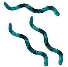
Bacteria are classified by their shape (morphology) and arrangement in colonies. Common shapes include:
Morphology | Description | Example Image |
|---|---|---|
Bacillus/Bacilli | Rod-shaped |
|
Coccus/Cocci | Round, spherical |
|
Spirillum/Spirilla | Spiral-shaped |
|


Bacterial arrangements include:
Arrangement | Description | Example Image |
|---|---|---|
Strepto- | Chains |
|
Staphylo- | Clusters |
|


Bacterial Staining: Gram Stain
Gram-Positive Bacteria: Thick peptidoglycan cell wall; retain crystal violet stain and appear purple.
Gram-Negative Bacteria: Thin peptidoglycan layer plus an outer membrane; do not retain crystal violet, stain red with safranin.
Medical Importance: Gram staining helps identify bacteria for appropriate treatment, as some antibiotics are effective only against specific types.
Viruses
Nature and Characteristics of Viruses
Viruses are not classified as prokaryotic or eukaryotic cells; they are acellular infectious agents. They require electron microscopes for visualization due to their small size (typically 20–300 nm). Viruses lack many characteristics of life, such as cellular organization, metabolism, and independent reproduction, but they do contain genetic material and can evolve.
Not Cells: Viruses are neither prokaryotic nor eukaryotic.
Microscopy: Electron microscopes are required to view viruses.
Size: Range from about 20 to 300 nanometers.
Characteristics Lacking: No cellular structure, metabolism, or independent reproduction.
Characteristics Present: Genetic material, ability to evolve, and reproduction (only within host cells).
Additional info: Viruses are important in medicine and biotechnology, both as pathogens and as tools for genetic engineering.
