 Back
BackMitosis and Meiosis: Cellular Division and Chromosome Dynamics
Study Guide - Smart Notes
 Tailored notes based on your materials, expanded with key definitions, examples, and context.
Cellular Division
Overview of Cellular Division
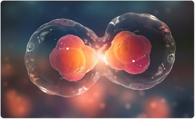
Cellular division is the process by which a parent cell divides to produce daughter cells. This process is fundamental for growth, development, repair, and reproduction in living organisms. There are three main types of cellular division: binary fission, mitosis, and meiosis.
Binary Fission: Occurs in prokaryotes (bacteria and archaea) and results in two genetically identical cells.
Mitosis: Responsible for growth, development, and repair in eukaryotes; produces two genetically identical diploid cells.
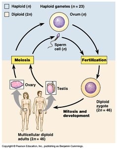
Meiosis: Produces gametes (sperm and egg) in sexually reproducing organisms; results in four genetically unique haploid cells.
Chromosome Structure and Ploidy
Chromosome Organization
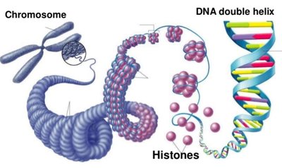
Chromosomes are highly organized structures composed of DNA and proteins. DNA wraps around histone proteins to form nucleosomes, which further coil to create chromatin and, ultimately, condensed chromosomes visible during cell division.
Nucleosome: The basic unit of DNA packaging, consisting of DNA wrapped around histone proteins.
Chromatin: The complex of DNA and proteins that forms chromosomes within the nucleus.
Chromosome: A single, long DNA molecule with associated proteins, carrying genetic information.
Ploidy: Diploid and Haploid Cells
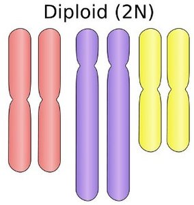
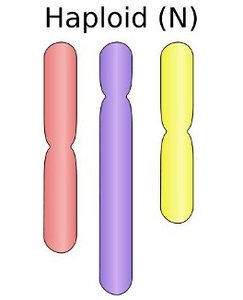
Ploidy refers to the number of sets of chromosomes in a cell. Most somatic (body) cells are diploid (2n), containing two sets of chromosomes, while gametes are haploid (n), containing one set.
Diploid (2n): Cells with two complete sets of chromosomes (e.g., human somatic cells have 46 chromosomes).
Haploid (n): Cells with one set of chromosomes (e.g., human gametes have 23 chromosomes).
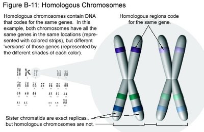
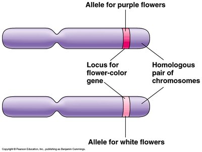
Homologous Chromosomes and Sex Chromosomes
Homologous chromosomes are pairs of chromosomes that are similar in size, shape, and gene content but may carry different alleles. Sex chromosomes determine the biological sex of an organism (e.g., X and Y in humans).
Homologous Chromosomes: Carry the same genes at the same loci but may have different alleles.
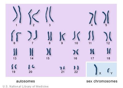
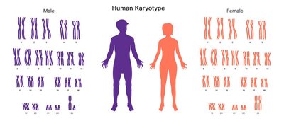
Sex Chromosomes: Chromosomes involved in sex determination (XX for females, XY for males in humans).
Karyotype Analysis
A karyotype is a visual representation of all the chromosomes in a cell, arranged by size, shape, and number. It is used to detect chromosomal abnormalities and determine sex.
Karyotype: A laboratory technique that produces an image of an individual's chromosomes.
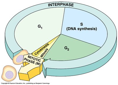
The Cell Cycle
Phases of the Cell Cycle
The cell cycle is the series of events that cells go through as they grow and divide. It consists of interphase (G1, S, G2) and the mitotic phase (mitosis and cytokinesis).
G1 Phase: Cell growth and normal functions.
S Phase: DNA replication (synthesis).
G2 Phase: Preparation for mitosis.
M Phase: Mitosis and cytokinesis.
Mitosis
Purpose and Overview
Mitosis is a type of nuclear division that results in two genetically identical diploid daughter cells. It is essential for growth, tissue repair, and asexual reproduction in some organisms. Mitosis is divided into five stages: prophase, prometaphase, metaphase, anaphase, and telophase, followed by cytokinesis.
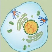
Prophase: Chromosomes condense, spindle fibers form, nuclear envelope breaks down.
Prometaphase: Kinetochores form, microtubules attach to chromosomes.
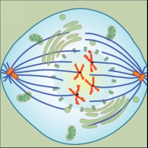
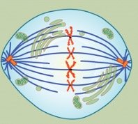
Metaphase: Chromosomes align at the metaphase plate.
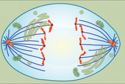
Anaphase: Sister chromatids separate and move to opposite poles.
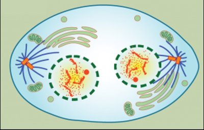
Telophase: Nuclear envelope reforms, chromosomes decondense.
Cytokinesis: Division of the cytoplasm, forming two daughter cells.
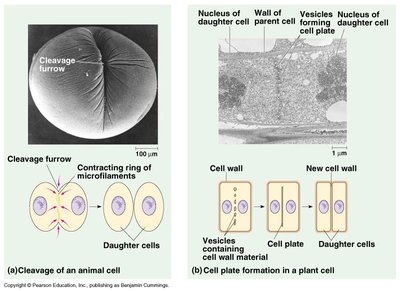
Cytokinesis
Cytokinesis is the process that divides the cytoplasm of a parental cell into two daughter cells. In animal cells, this occurs via cleavage furrow formation, while in plant cells, a cell plate forms to separate the two new cells.
Meiosis
Purpose and Overview
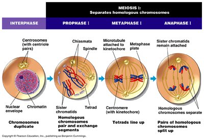
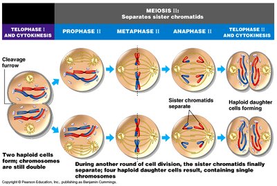
Meiosis is a specialized form of cell division that reduces the chromosome number by half, producing four genetically unique haploid gametes. It consists of two sequential divisions: meiosis I and meiosis II. Meiosis introduces genetic diversity through crossing over and independent assortment.
Meiosis I: Homologous chromosomes separate, reducing chromosome number by half.
Meiosis II: Sister chromatids separate, similar to mitosis.

Stages of Meiosis
Each meiotic division is subdivided into prophase, metaphase, anaphase, and telophase. Key events include synapsis and crossing over during prophase I, and the separation of homologous chromosomes (meiosis I) and sister chromatids (meiosis II).
Prophase I: Homologous chromosomes pair and exchange genetic material (crossing over).
Metaphase I: Homologous pairs align at the metaphase plate.
Anaphase I: Homologous chromosomes separate to opposite poles.
Telophase I and Cytokinesis: Two haploid cells form.
Meiosis II: Similar to mitosis; sister chromatids separate, resulting in four haploid cells.
Genetic Variation: Crossing Over
Crossing over occurs during prophase I of meiosis, where homologous chromosomes exchange genetic material. This process increases genetic diversity in offspring.
Comparison of Mitosis and Meiosis
Feature | Mitosis | Meiosis |
|---|---|---|
Number of divisions | 1 | 2 |
Number of daughter cells | 2 | 4 |
Chromosome number in daughter cells | Diploid (2n) | Haploid (n) |
Genetic identity | Identical to parent | Genetically unique |
Role | Growth, repair, asexual reproduction | Sexual reproduction (gamete formation) |
Applications and Laboratory Observations
Microscopic examination of dividing cells (e.g., onion root tip, whitefish blastula, grasshopper testes) allows visualization of mitosis and meiosis stages. Laboratory simulations (e.g., using Lego kits) help reinforce understanding of chromosome behavior during division.
Example: In humans, meiosis occurs in the testes (males) and ovaries (females), producing sperm and eggs, respectively. Fertilization restores the diploid chromosome number in the zygote.
Additional info: The duration of meiosis varies: about one month in males and 10–50 years in females, due to the arrest of oocytes in prophase I until ovulation.
