 Back
BackMitosis and the Eukaryotic Cell Cycle: Structure, Phases, and Regulation
Study Guide - Smart Notes
 Tailored notes based on your materials, expanded with key definitions, examples, and context.
Mitosis and the Cell Cycle
Overview of the Cell Cycle
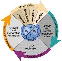
The cell cycle is the series of events that cells go through as they grow and divide. It consists of interphase (G1, S, G2) and the mitotic (M) phase. The cycle ensures that genetic material is accurately replicated and distributed to daughter cells.
G1 phase: Cell grows and performs normal metabolic roles.
S phase: DNA replication occurs, resulting in duplicated chromosomes.
G2 phase: Further growth and preparation for mitosis.
M phase (Mitosis): Division of the nucleus and cytoplasm to form two daughter cells.
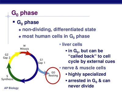
G0 phase: A resting, non-dividing state; many differentiated cells (e.g., nerve and muscle cells) remain here permanently.
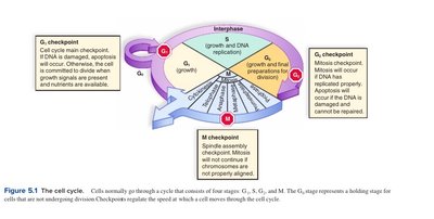
Cell Cycle Checkpoints
Checkpoints are regulatory points where the cell examines internal and external cues to decide whether to proceed with division. The main checkpoints are:
G1 checkpoint: Checks for DNA damage and sufficient resources. If conditions are not met, the cell may enter G0.
G2 checkpoint: Ensures DNA replication is complete and undamaged before mitosis.
M checkpoint (Spindle checkpoint): Ensures all chromosomes are properly attached to the spindle before anaphase.
Phases of Mitosis
Introduction to Mitosis
Mitosis is the process by which a eukaryotic cell separates its duplicated chromosomes into two identical sets, resulting in two genetically identical daughter cells. It is conventionally divided into five phases:
Prophase
Prometaphase
Metaphase
Anaphase
Telophase
Cytokinesis (division of the cytoplasm) usually overlaps with telophase.
G2 of Interphase
Before mitosis begins, the cell is in G2 of interphase:
Chromosomes have been replicated but are not yet condensed.
Nucleolus is present and active in ribosome production.
Nuclear envelope surrounds the nucleus.
Two centrosomes (in animal cells) organize the mitotic spindle.
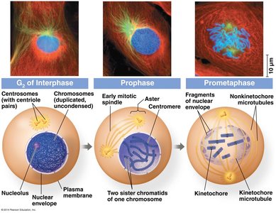
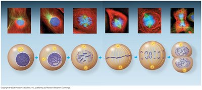
Prophase
During prophase:
Chromatin condenses into visible chromosomes, each with two sister chromatids.
The mitotic spindle begins to form from centrosomes.
Asters (radial microtubule arrays) form at centrosomes.
Centrosomes move apart.
Nucleoli disappear.

Prometaphase
During prometaphase:
Nuclear envelope fragments, allowing spindle microtubules to access chromosomes.
Each chromatid develops a kinetochore at its centromere.
Some spindle microtubules attach to kinetochores, others interact with microtubules from the opposite pole.

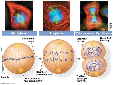
Metaphase
During metaphase:
Spindle is fully formed, with centrosomes at opposite poles.
Chromosomes align at the metaphase plate (equidistant between poles).
Kinetochores of sister chromatids are attached to microtubules from opposite poles.


Anaphase
During anaphase:
Sister chromatids separate and move toward opposite poles, becoming individual chromosomes.
Kinetochore microtubules shorten, pulling chromosomes centromere-first.
Nonkinetochore microtubules lengthen, elongating the cell.
Telophase and Cytokinesis
During telophase:
Two daughter nuclei form, and nuclear envelopes reappear.
Chromosomes decondense back into chromatin.
Spindle microtubules depolymerize.
Cytokinesis divides the cytoplasm, resulting in two genetically identical daughter cells.

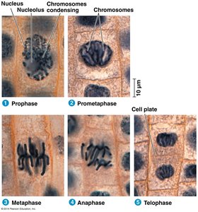
Observing Mitosis
Scientists observe mitosis by staining DNA and using microscopy to visualize the different stages. This allows for the identification of each phase based on chromosome and spindle appearance.
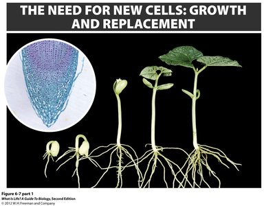
Cell Division in Plants and Animals
Plant Cell Division
In plants, cell division is especially rapid at the root tip, which is a key site for growth and development. Plant mitosis is similar to animal mitosis, but cytokinesis involves the formation of a cell plate.
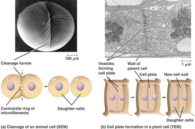
Animal Cell Division
In animal cells, cytokinesis occurs via cleavage, where a contractile ring pinches the cell into two daughter cells.
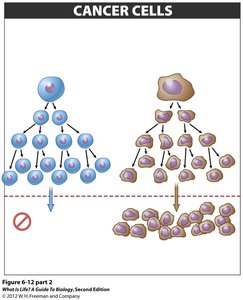
Regulation and Abnormal Cell Division
Cancer Cells vs. Normal Cells
Normal somatic cells can divide only a limited number of times due to regulatory mechanisms. In contrast, cancer cells lose this regulation and can divide indefinitely, leading to tumor formation and uncontrolled growth.
Normal cells: Undergo senescence and stop dividing after a certain number of divisions.
Cancer cells: Continue to divide without limit, often due to mutations in genes controlling the cell cycle.
Summary Table: Mitosis Phases and Key Events
Phase | Main Events |
|---|---|
Prophase | Chromosomes condense, spindle forms, nucleolus disappears |
Prometaphase | Nuclear envelope fragments, kinetochores form, spindle attaches |
Metaphase | Chromosomes align at metaphase plate |
Anaphase | Sister chromatids separate, move to opposite poles |
Telophase | Nuclear envelopes reform, chromosomes decondense |
Cytokinesis | Cytoplasm divides, two daughter cells form |
Additional info:
Some images and explanations were expanded for clarity and completeness based on standard biology curriculum.
Key terms such as chromatin, centrosome, kinetochore, and metaphase plate are defined in context.
Equations are not directly relevant to mitosis, but the process is tightly regulated by molecular checkpoints and protein complexes (e.g., cyclins and CDKs).
