 Back
Mitosis, the Cell Cycle, and Cancer: Regulation and Consequences of Cell Division
Study Guide - Smart Notes
 Tailored notes based on your materials, expanded with key definitions, examples, and context.
Mitosis and the Cell Cycle
Introduction to Cell Division
Cell division is a fundamental biological process by which a parent cell divides to produce new daughter cells. This process is essential for growth, tissue repair, and reproduction in all living organisms. In multicellular organisms, cell division enables development from a single cell and maintains tissue homeostasis throughout life.
Parent Cell: The original cell that undergoes division.
Daughter Cells: The two new cells produced from the division of the parent cell.
Purposes of Cell Division:
Growth of multicellular organisms
Repair of damaged tissues
Reproduction (asexual via mitosis, sexual via meiosis)
The Cell Cycle: Overview and Phases
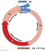
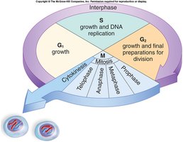
The cell cycle is an ordered sequence of events that leads to cell division and the production of two genetically identical daughter cells. It consists of two main stages: Interphase (preparation) and M phase (mitosis and cytokinesis).
Interphase: The cell grows, replicates its DNA, and prepares for division. It includes three subphases:
G1 phase (First Gap): Cell growth and normal functions; organelles double and proteins for DNA synthesis are produced.
S phase (Synthesis): DNA replication; chromosomes are duplicated.
G2 phase (Second Gap): Preparation for division; proteins needed for mitosis are synthesized.
M phase (Mitotic phase): Division of the nucleus (mitosis) and cytoplasm (cytokinesis).
Genetic Material and Chromosome Terminology
During the cell cycle, the structure and organization of genetic material change to ensure accurate DNA replication and segregation.
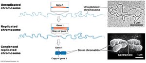
Chromosome: A single, long DNA molecule with associated proteins (chromatin in eukaryotes).
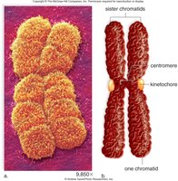
Chromatid: Each of the two identical copies of a duplicated chromosome.
Sister Chromatids: The two chromatids of a duplicated chromosome, joined at the centromere.
Centromere: The region where sister chromatids are joined and where spindle fibers attach during mitosis.
Kinetochore: Protein complexes on either side of the centromere; attachment site for spindle microtubules.
Mitosis and Cytokinesis
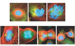
Phases of Mitosis
Mitosis is the process by which a eukaryotic cell separates its duplicated chromosomes into two identical sets. It is followed by cytokinesis, the division of the cytoplasm.
Prophase: Chromosomes condense, spindle apparatus forms.
Prometaphase: Nuclear envelope breaks down, spindle fibers attach to kinetochores.
Metaphase: Chromosomes align at the metaphase plate.
Anaphase: Sister chromatids separate and move toward opposite poles.
Telophase: Nuclear envelopes reform around the two sets of chromosomes.
Cytokinesis: Division of the cytoplasm, resulting in two daughter cells.
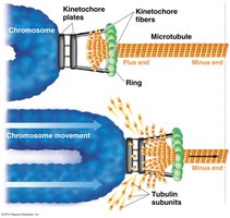
Chromosome Movement During Mitosis
Chromosome movement is driven by the shortening of microtubules attached to kinetochores, facilitated by motor proteins.
Microtubules depolymerize at the kinetochore, pulling chromatids apart.
Motor proteins "walk" along microtubules, aiding chromosome movement.
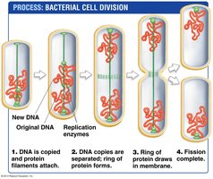
Prokaryotic Cell Division: Binary Fission
Prokaryotes divide by binary fission, a simpler and faster process than mitosis.
DNA is replicated, and each copy attaches to the cell membrane.
The cell elongates, and the plasma membrane pinches inward, dividing the cell.
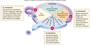
Regulation of the Cell Cycle
Cell Cycle Control and Checkpoints
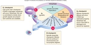
Cell division is tightly regulated by checkpoints that ensure the fidelity of cell division and prevent uncontrolled proliferation.
G1 Checkpoint: Checks for cell size, nutrients, growth factors, and DNA damage.
G2 Checkpoint: Ensures DNA replication is complete and undamaged; checks for activated MPF.
Metaphase (Spindle) Checkpoint: Ensures all chromosomes are properly attached to the spindle apparatus.
G0 Phase: Non-dividing state where cells perform normal functions but do not prepare for division.
Mitosis Promoting Factor (MPF) and Cyclin Regulation
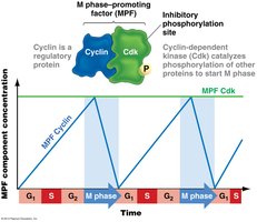
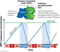
The transition from G2 to M phase is regulated by the Mitosis Promoting Factor (MPF), a complex of cyclin and cyclin-dependent kinase (Cdk).
Cyclin: Regulatory protein whose concentration fluctuates during the cell cycle.
Cdk: Enzyme that is active only when bound to cyclin; phosphorylates target proteins to initiate mitosis.
MPF activity is regulated by cyclin concentration and phosphorylation status.
Apoptosis: Programmed Cell Death
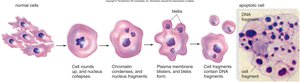
Cells with irreparable DNA damage undergo apoptosis, a controlled process of cell death that prevents the propagation of defective cells.
Prevents tumorigenesis, shapes tissues during development, and limits viral spread.
Characterized by nuclear breakdown, membrane blebbing, and DNA fragmentation.
Cancer: Loss of Cell Cycle Control
Characteristics and Causes of Cancer
Cancer is a group of diseases characterized by uncontrolled cell division due to the breakdown of cell cycle regulation. Multiple mutations in genes that control the cell cycle are required for cancer to develop.
Proto-oncogenes: Genes that promote cell division; mutations can convert them to oncogenes, driving cancer.
Tumor Suppressor Genes: Genes that inhibit cell division or promote apoptosis; loss-of-function mutations remove these brakes.
Mutagens: Physical or chemical agents (e.g., radiation, carcinogens) that cause DNA mutations.
Viruses: Some viruses can disrupt cell cycle regulation, leading to cancer (e.g., HPV, Epstein-Barr virus).
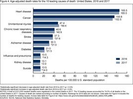
Hallmarks of Cancer Cells
Cancer cells exhibit several key features that distinguish them from normal cells:
Divide without growth factors
Ignore growth-inhibiting signals (loss of contact inhibition)
Evade apoptosis
Divide indefinitely (immortalized)
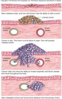
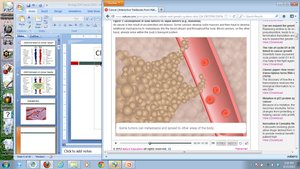
Metastasize (invade other tissues)
Promote angiogenesis (blood vessel growth)
Genetic and Environmental Factors in Cancer
Cancer development is multifactorial, involving genetic predisposition, environmental exposures, and lifestyle choices.
High-fat diets, smoking, and exposure to mutagens increase cancer risk.
Incidence increases with age due to accumulation of mutations (multiple hit hypothesis).
Summary Table: Key Genes in Cell Cycle Regulation
Gene Type | Normal Function | Effect of Mutation |
|---|---|---|
Proto-oncogene | Promotes cell division | Oncogene drives uncontrolled division |
Tumor Suppressor | Inhibits cell division, promotes apoptosis | Loss of function removes cell cycle brakes |
Multiple Hit Hypothesis
At least four mutations in cell cycle control genes are typically required for a cell to become cancerous. DNA repair mechanisms usually prevent rapid accumulation of these mutations, explaining why cancer risk increases with age.
Additional info: The cell cycle and its regulation are central to understanding both normal cellular function and the development of diseases such as cancer. Mastery of these concepts is essential for further study in cell biology, genetics, and medicine.
