 Back
BackMitotic Division and Regulation of the Eukaryotic Cell Cycle
Study Guide - Smart Notes
 Tailored notes based on your materials, expanded with key definitions, examples, and context.
Mitotic Division
Overview of Mitosis
Mitosis is the process by which a single cell divides to produce two genetically identical daughter cells. This process is essential for growth, development, and tissue repair in multicellular organisms, and for asexual reproduction in unicellular organisms.
Purpose: Development, growth, and replacement of damaged or old cells.
Genetic Consistency: Daughter cells are genetically identical to the parent cell.
Phases of the Cell Cycle
The cell cycle consists of interphase and the mitotic (M) phase. Interphase is subdivided into three phases: G1 (first gap), S (synthesis), and G2 (second gap). Chromosome duplication occurs only during the S phase.
G1 Phase: Cell growth and preparation for DNA replication.
S Phase: DNA synthesis and chromosome duplication.
G2 Phase: Further growth and preparation for mitosis.
Stages of Mitosis
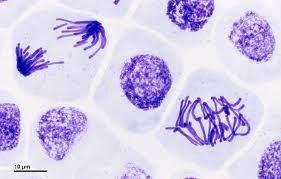
Mitosis is divided into five main stages: prophase, prometaphase, metaphase, anaphase, and telophase. Each stage is characterized by specific events involving the chromosomes and the mitotic spindle.
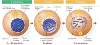
Prophase: Chromosomes condense, spindle apparatus begins to form, and nucleolus disappears.
Prometaphase: Nuclear envelope fragments, spindle microtubules attach to kinetochores.
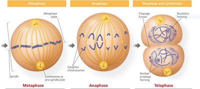
Metaphase: Chromosomes align at the metaphase plate.
Anaphase: Sister chromatids separate and move toward opposite poles.
Telophase: Nuclear envelopes reform, chromosomes decondense.
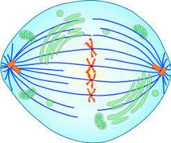
The Mitotic Spindle
The mitotic spindle is a structure composed of microtubules that orchestrates the movement of chromosomes during mitosis. In animal cells, spindle assembly begins at the centrosomes, which replicate and migrate to opposite poles of the cell.
Centrosome: Microtubule-organizing center.
Aster: Radial array of short microtubules extending from each centrosome.
Spindle Microtubules: Attach to kinetochores and facilitate chromosome movement.
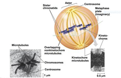
Chromosome Movement and Separation
During prometaphase, spindle microtubules attach to kinetochores, protein complexes at the centromeres. At metaphase, chromosomes align at the metaphase plate. In anaphase, cohesins are cleaved, allowing sister chromatids to separate and move toward opposite poles, primarily through the action of motor proteins and microtubule depolymerization (the "Pac-man" mechanism).
Kinetochore: Protein complex at the centromere where spindle fibers attach.
Metaphase Plate: Imaginary plane equidistant from the spindle poles where chromosomes align.
Pac-man Mechanism: Chromosomes are moved as microtubules depolymerize at the kinetochore ends.
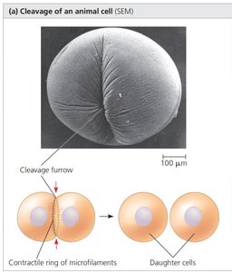
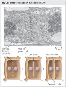
Cytokinesis
Cytokinesis is the division of the cytoplasm, which usually begins during telophase. The process differs between animal and plant cells:
Animal Cells: Formation of a cleavage furrow, which pinches the cell in two.
Plant Cells: Formation of a cell plate from Golgi-derived vesicles, which develops into a new cell wall.
Cell Division in Prokaryotes and Other Eukaryotes
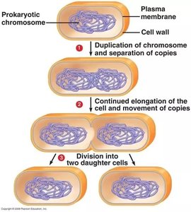
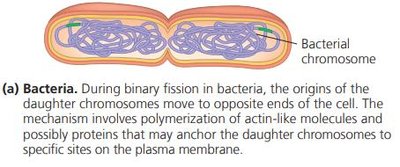
Binary Fission in Bacteria
Bacteria reproduce by binary fission, a process in which the chromosome is duplicated and the cell divides into two genetically identical daughter cells.
Steps: Chromosome duplication, elongation of the cell, and division into two cells.
Mechanism: Involves actin-like molecules and proteins anchoring chromosomes to the plasma membrane.
Variations in Eukaryotic Cell Division
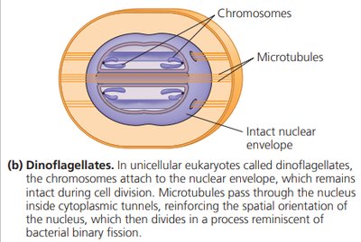
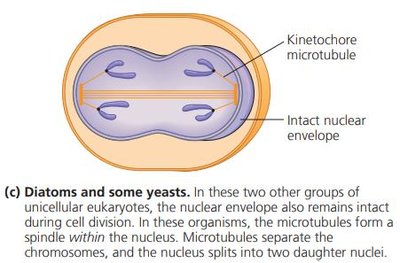
Some unicellular eukaryotes, such as dinoflagellates, diatoms, and yeasts, exhibit variations in the process of cell division, particularly in the behavior of the nuclear envelope and spindle formation.
Dinoflagellates: Chromosomes attach to the nuclear envelope, which remains intact; microtubules pass through cytoplasmic tunnels.
Diatoms and Yeasts: Nuclear envelope remains intact; spindle forms within the nucleus.
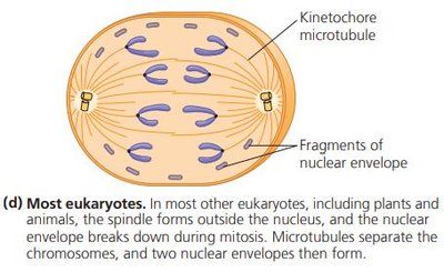
Most Eukaryotes: Spindle forms outside the nucleus; nuclear envelope breaks down during mitosis.
Regulation of the Eukaryotic Cell Cycle
Molecular Control of the Cell Cycle
The frequency of cell division varies with cell type and is regulated at the molecular level. Checkpoints in the cell cycle ensure that critical processes are completed before the cell proceeds to the next stage.
Checkpoints: Control points where stop and go-ahead signals regulate the cycle.
Growth Factors: External signals that stimulate cell division.
Loss of Cell Cycle Control and Cancer
Cancer cells escape normal cell cycle controls, leading to uncontrolled division. They may produce their own growth factors, signal without external growth factors, or have abnormal control systems. Cells that divide indefinitely undergo transformation and may form tumors.
Benign Tumor: Abnormal cells remain at the original site.
Malignant Tumor: Invades surrounding tissues and can metastasize to other parts of the body.
Treatment: Localized tumors may be treated with radiation; metastatic cancers are treated with chemotherapies targeting the cell cycle.
Personalized Medicine: Advances in cell cycle understanding and DNA sequencing are leading to more personalized cancer treatments.
