 Back
BackMolecular Biology Lab: DNA, Restriction Enzymes, and Gel Electrophoresis
Study Guide - Smart Notes
 Tailored notes based on your materials, expanded with key definitions, examples, and context.
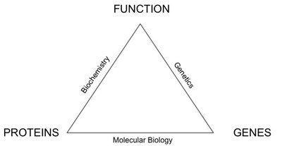
Molecular Biology Overview
Introduction to Molecular Biology
Molecular biology is the branch of biology that deals with the molecular basis of biological activity, focusing on the structure and function of genes and gene expression. It integrates concepts from genetics and biochemistry to understand how genetic information is encoded, replicated, and expressed within cells.
Genes: Segments of DNA that code for proteins or functional RNA molecules.
Gene Expression: The process by which information from a gene is used to synthesize a functional gene product, often a protein.
Key Techniques: Molecular cloning, Polymerase Chain Reaction (PCR), gel electrophoresis, restriction enzyme digestion, blotting and probing, and DNA microarrays.
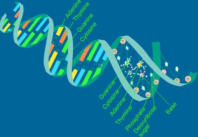
DNA Structure and Properties
Nucleic Acids and DNA Composition
DNA (deoxyribonucleic acid) is the hereditary material in almost all living organisms. It is composed of nucleotides, each containing a phosphate group, a deoxyribose sugar, and a nitrogenous base.
Nitrogenous Bases: Purines (adenine [A], guanine [G]) and pyrimidines (thymine [T], cytosine [C]).
Phosphate Backbone: Gives DNA its negative charge.
Double Helix: Two strands of DNA wind around each other, held together by hydrogen bonds between complementary bases (A-T, G-C).
Laboratory Techniques in Molecular Biology
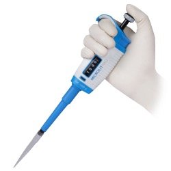
Proper Pipetting Technique
Accurate pipetting is essential for molecular biology experiments to ensure precise measurement and transfer of small liquid volumes.
Use a micropipette with disposable tips.
Always pre-wet the tip and pipette at a consistent angle.
Avoid air bubbles and ensure proper tip ejection after use.
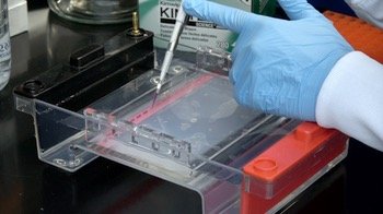
Loading Electrophoresis Gels
Loading samples into an agarose gel is a critical step in gel electrophoresis, allowing for the separation of DNA fragments by size.
Use a micropipette to carefully load DNA samples into wells in the gel.
Avoid puncturing the gel or mixing samples between wells.
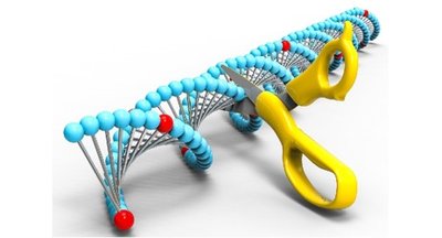
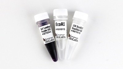
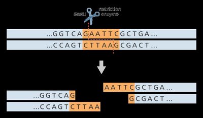
Restriction Enzymes
Restriction enzymes are proteins that cut DNA at specific nucleotide sequences, generating fragments with defined ends. They are essential tools for DNA analysis and recombinant DNA technology.
Recognition Sites: Short, specific DNA sequences where the enzyme cuts.
Sticky Ends: Overhanging single-stranded DNA produced by staggered cuts, useful for ligating DNA fragments.
Blunt Ends: Straight cuts with no overhangs.
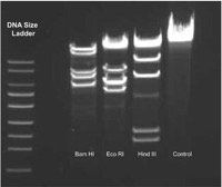
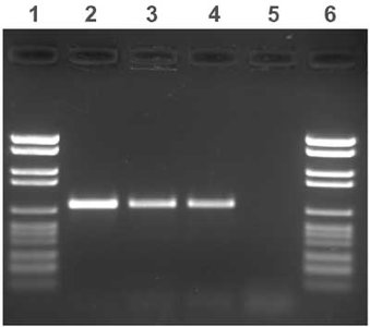
Gel Electrophoresis
Gel electrophoresis is a technique used to separate DNA fragments by size using an electric field. DNA, being negatively charged, migrates toward the positive electrode through a porous agarose matrix.
Smaller Fragments: Move faster and travel farther through the gel.
DNA Ladder: A mixture of DNA fragments of known sizes used as a reference to estimate the size of sample fragments.
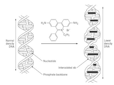
Visualization: DNA is stained with ethidium bromide, which intercalates between bases and fluoresces under UV light.
Ethidium Bromide and DNA Visualization
Ethidium bromide is a fluorescent dye used to visualize DNA in gels. It binds to DNA by intercalating between base pairs, but it is a mutagen and must be handled with care.
Visualization: Glows under UV light, making DNA bands visible.
Hazards: Can cause mutations and is potentially carcinogenic.
Experimental Design: Restriction Digests and Gel Analysis
Restriction Digest Experiment
In a typical restriction digest experiment, DNA (such as a plasmid) is incubated with one or more restriction enzymes to cut the DNA at specific sites. The resulting fragments are then separated and analyzed by gel electrophoresis.
Single enzyme digestion (e.g., EcoRI or SalI) produces a specific number and size of DNA fragments.
Double digestion (e.g., EcoRI and SalI together) produces a different pattern, useful for mapping restriction sites.
Expected results depend on the number and location of recognition sites in the DNA.
Interpreting Gel Electrophoresis Results
After electrophoresis, the pattern of DNA bands is compared to the DNA ladder to determine the size of fragments. The number and size of bands provide information about the DNA and the effectiveness of the restriction digest.
One band: Uncut circular plasmid or linearized DNA.
Two or more bands: DNA cut at one or more sites.
Band sizes are estimated by comparing to the DNA ladder.
Laboratory Safety
Safe Handling of Chemicals and Equipment
Proper laboratory safety is essential when working with hazardous chemicals like ethidium bromide and when using electrical equipment for gel electrophoresis.
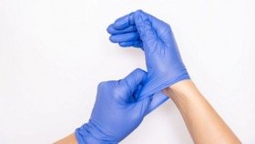
Always wear gloves and lab coats.
Handle ethidium bromide only at designated gel benches.
Do not heat ethidium bromide or touch other surfaces with contaminated gloves.
Dispose of all hazardous waste according to lab protocols.
Summary Table: Key Techniques and Their Purposes
Technique | Main Purpose | Key Reagents/Equipment |
|---|---|---|
Pipetting | Accurate measurement and transfer of liquids | Micropipette, tips |
Restriction Digest | Cut DNA at specific sequences | Restriction enzymes (e.g., EcoRI, SalI) |
Gel Electrophoresis | Separate DNA fragments by size | Agarose gel, electrophoresis chamber, power supply, DNA ladder |
DNA Visualization | Detect DNA bands in gel | Ethidium bromide, UV transilluminator |
Practice Questions for Lab Preparation
How many DNA bands do you expect, and what sizes, after cutting a plasmid with EcoRI, SalI, or both?
How does the number of restriction sites affect the banding pattern?
Why is it important to use a DNA ladder in gel electrophoresis?
What safety precautions must be taken when handling ethidium bromide?
Additional info: This guide covers core molecular biology lab techniques relevant to DNA analysis, including restriction enzyme digestion and gel electrophoresis, as well as essential safety practices. These concepts are foundational for understanding gene structure, function, and manipulation in modern biology.
