 Back
BackMolecular Biology of the Gene: DNA Structure and Replication
Study Guide - Smart Notes
 Tailored notes based on your materials, expanded with key definitions, examples, and context.
Molecular Biology of the Gene
Structure of DNA
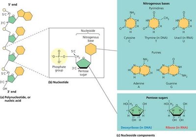
The structure of DNA is fundamental to understanding its function in genetic inheritance and cellular processes. DNA is a polymer composed of nucleotides, each consisting of a pentose sugar, a phosphate group, and a nitrogenous base.
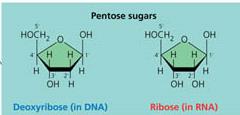
Nucleotide Structure: Each nucleotide contains a pentose sugar (deoxyribose in DNA, ribose in RNA), a phosphate group, and a nitrogenous base.
Pentose Sugars: Deoxyribose (in DNA) lacks an oxygen atom on the 2' carbon compared to ribose (in RNA).
Nitrogenous Bases: DNA contains adenine (A), guanine (G), cytosine (C), and thymine (T). RNA contains uracil (U) instead of thymine.
Numbering of Atoms: Atoms in the sugar are numbered with primes (e.g., 2', 3', 5') to distinguish them from atoms in the nitrogenous base.
Attachment Points: The nitrogenous base attaches to the 1' carbon, the phosphate group to the 5' carbon, and the 3' carbon forms a bond with the next nucleotide.
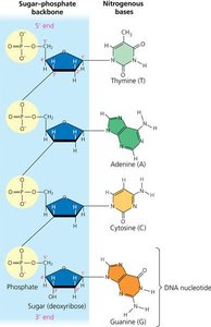
DNA Strand Directionality and Backbone
DNA strands have directionality due to the orientation of the sugar-phosphate backbone. The two ends of a DNA strand are chemically distinct:
5' End: Has a phosphate group attached to the 5' carbon of the sugar.
3' End: Has a hydroxyl group attached to the 3' carbon.
Phosphodiester Bonds: Adjacent nucleotides are joined by covalent bonds between the 3' hydroxyl of one nucleotide and the 5' phosphate of the next, forming a sugar-phosphate backbone.
Antiparallel Strands: The two DNA strands run in opposite directions (one 5'→3', the other 3'→5').
Base Sequence and Genetic Information
The sequence of nitrogenous bases along a DNA strand encodes genetic information. Each gene has a unique sequence, which determines the amino acid sequence of proteins.
Genetic Code: The order of bases specifies the primary structure of proteins, which determines their three-dimensional structure and function.
Limitless Combinations: With hundreds to thousands of nucleotides per gene, the possible base sequences are effectively limitless.
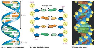
Double Helix Structure
DNA exists as a double helix, with two antiparallel strands held together by hydrogen bonds between complementary bases (A-T and G-C).
Complementary Base Pairing: Adenine pairs with thymine (A-T), and guanine pairs with cytosine (G-C).
Helical Structure: The double helix has a uniform diameter and specific spacing between base pairs.
DNA Replication
Overview of DNA Replication
DNA replication is the process by which a cell copies its DNA before cell division. It is semi-conservative, meaning each daughter DNA molecule consists of one parental and one newly synthesized strand.
Origins of Replication: Replication begins at specific sites called origins of replication.
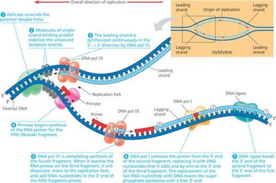
Replication Forks: Helicase unwinds the DNA, forming replication forks where new strands are synthesized.
Single-Strand Binding Proteins: These proteins stabilize the unwound DNA and prevent the strands from re-annealing.
Primase: Synthesizes short RNA primers to provide a starting point for DNA polymerase.
DNA Polymerase: Adds nucleotides to the 3' end of the primer, synthesizing new DNA in the 5'→3' direction.

Leading and Lagging Strands
Because DNA polymerase can only add nucleotides to the 3' end, replication differs between the two template strands:
Leading Strand: Synthesized continuously toward the replication fork.
Lagging Strand: Synthesized discontinuously away from the fork in short segments called Okazaki fragments.
Okazaki Fragments: Each fragment begins with an RNA primer; DNA polymerase extends the fragment, and DNA ligase joins the fragments into a continuous strand.
Replication Bubbles and Semi-Conservative Model
In eukaryotes, replication occurs at multiple origins, forming replication bubbles that expand until they merge, resulting in two complete DNA molecules.
Semi-Conservative Replication: Each daughter DNA molecule contains one old (parental) strand and one new strand.
Replication Accuracy: DNA polymerase proofreads and corrects errors, resulting in high fidelity (only 1 error per billion nucleotides).

Telomeres and Telomerase
Structure and Function of Telomeres
Telomeres are repetitive nucleotide sequences at the ends of eukaryotic chromosomes that protect genetic information from degradation during replication.
Sequence: In humans, the telomere repeat is 5' TTAGGG 3'.
Function: Telomeres prevent the loss of vital genetic information during successive rounds of DNA replication.
Species Variation: Telomere sequences differ among species.
Group | Organism | Telomeric Repeat (5' to 3') |
|---|---|---|
Vertebrates | Human, mouse | TTAGGG |
Filamentous fungi | Neurospora crassa | TTAGGG |
Green algae | Chlamydomonas | TTTTAGGG |
Roundworms | Ascaris lumbricoides | TTAGGC |
Insects | Bombyx mori | TTAGG |
Budding yeasts | Candida albicans | GGTGTACGGATGTCTAACTTCTT |
Telomere Shortening and the End Replication Problem
During DNA replication, the lagging strand cannot be fully replicated at the ends, leading to progressive telomere shortening with each cell division.
End Replication Problem: The inability of DNA polymerase to replicate the very end of linear chromosomes results in the loss of telomeric DNA.
Cellular Aging: Human telomeres lose about 100 base pairs per cell division, limiting the number of times a cell can divide (the Hayflick limit).
Non-dividing Cells: Telomeres do not shorten in cells that do not divide, such as heart muscle cells.
Role of Telomerase
Telomerase is an enzyme that extends telomeres in germ cells, allowing for complete replication of chromosome ends and maintaining genetic integrity in gametes.
Function: Adds telomeric repeats to the 3' end of DNA, enabling DNA polymerase to finish replication.
Activity: Active in germ cells, not in most somatic cells; ensures zygotes have full-length telomeres.
Telomeres and Cloning
Cloning from adult cells can result in organisms with shorter telomeres, as seen in Dolly the sheep, raising questions about the implications for aging and cellular health in clones.
Example: Dolly's telomeres were only 80% as long as those in a normal one-year-old sheep, likely due to the donor cell's history of divisions without telomerase activity.
