 Back
BackMotor Mechanisms and Photosynthesis: Structure, Function, and Regulation in Biology
Study Guide - Smart Notes
 Tailored notes based on your materials, expanded with key definitions, examples, and context.
Motor Mechanisms in Animals
The Vertebrate Nervous System and Motor Control
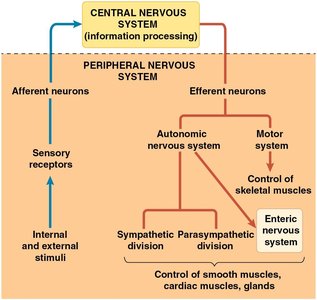
The vertebrate nervous system is organized to process information and coordinate movement. The central nervous system (CNS) integrates information, while the peripheral nervous system (PNS) transmits signals between the CNS and the rest of the body. The motor system carries signals to skeletal muscles, enabling voluntary and involuntary movements.
Motor neurons (efferent neurons) communicate with muscle cells at specialized synapses called neuromuscular junctions (NMJs).
At the NMJ, the neurotransmitter acetylcholine (ACh) is released, binding to ligand-gated ion channels on the muscle cell, initiating muscle contraction.
Types of Muscle in Vertebrates
Vertebrates possess three main types of muscle tissue, each with distinct structure and function:
Cardiac muscle: Found in the heart, responsible for pumping blood; involuntary control.
Smooth muscle: Located in organs, moves substances like blood and food; involuntary control.
Skeletal muscle: Attached to bones, responsible for locomotion; voluntary control.
All muscle types contain actin and myosin filaments, which are essential for contraction.
Structure of Vertebrate Skeletal Muscle
Skeletal muscle is organized from macroscopic to microscopic levels:
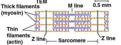
A muscle consists of bundles of muscle fibers (cells), each containing multiple nuclei.
Within each muscle fiber are myofibrils, which are bundles of contractile protein filaments.
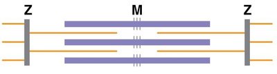
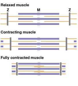
Myofibrils are composed of repeating units called sarcomeres, the basic contractile units of muscle.

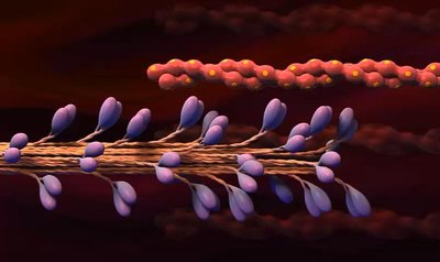
Protein Filaments and the Sliding Filament Model
Muscle contraction is driven by the interaction of two types of protein filaments:
Thin filaments: Composed mainly of actin, arranged in two coiled strands.
Thick filaments: Composed of myosin, with heads that bind to actin.
During contraction, these filaments slide past each other, shortening the sarcomere but not the individual filaments.
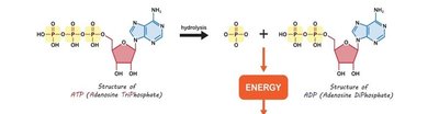
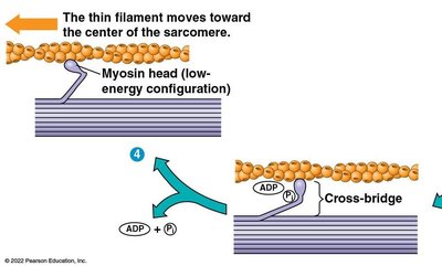
The Crossbridge Cycle and ATP
The crossbridge cycle describes the repeated binding and release of myosin heads to actin filaments, powered by ATP hydrolysis:
ATP binds to myosin, causing it to detach from actin.
ATP is hydrolyzed to ADP and Pi, energizing the myosin head.
Myosin binds to actin, forming a crossbridge.
Release of ADP and Pi triggers the power stroke, pulling actin past myosin.
Cycle repeats as new ATP binds.
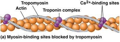
Regulation of Muscle Contraction by Calcium Ions
Muscle contraction is regulated by the presence of calcium ions (Ca2+) and associated regulatory proteins:
At rest, tropomyosin blocks myosin-binding sites on actin.
When Ca2+ binds to troponin, tropomyosin shifts, exposing binding sites for myosin.
Ca2+ is released from the sarcoplasmic reticulum in response to nerve impulses.
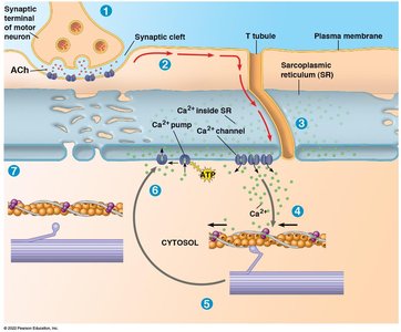
Muscle Relaxation
Muscle relaxation is a passive process that occurs when neural stimulation ceases:
Motor neuron input stops, acetylcholine release ends, and Ca2+ is pumped back into the sarcoplasmic reticulum.
Tropomyosin re-covers the myosin-binding sites, and the muscle relaxes.

Types of Skeletal Muscle Fibers
Skeletal muscles contain different fiber types adapted for specific functions:
Slow-twitch (oxidative) fibers: Contract slowly, resist fatigue, rely on aerobic respiration, and have high myoglobin content.
Fast-twitch fibers: Contract rapidly, fatigue quickly, and can be oxidative (aerobic) or glycolytic (anaerobic).
Slow-Twitch Oxidative | Fast-Twitch Oxidative | Fast-Twitch Glycolytic | |
|---|---|---|---|
Contraction speed | Slow | Fast | Fast |
Major ATP source | Aerobic respiration | Aerobic respiration | Glycolysis |
Rate of fatigue | Slow | Intermediate | Fast |
Mitochondria | Many | Many | Few |
Myoglobin content | High (red muscle) | High (red muscle) | Low (white muscle) |
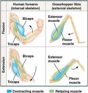
Animal Skeletons and Locomotion
Skeletons provide structure, support, and protection, and serve as attachment points for muscles. There are three main types of skeletons:
Hydrostatic skeletons: Fluid-filled compartments (e.g., cnidarians, annelids).
Exoskeletons: Hard external coverings (e.g., arthropods, molluscs).
Endoskeletons: Internal skeletons (e.g., vertebrates).
Forms and Energetics of Locomotion
Locomotion requires energy to overcome friction and gravity. The medium (land, water, air) influences movement strategies and energy costs:
Land locomotion: Must overcome gravity; examples include walking, running, hopping.
Water locomotion: Must overcome friction; streamlined bodies and various swimming methods are adaptations.
Air locomotion: Must overcome both gravity and friction; adaptations include wings, reduced body mass, and air-filled bones.

The energy cost of locomotion can be estimated by measuring oxygen consumption or carbon dioxide production. Generally, swimming is the most energy-efficient mode, followed by flying and then running.
Photosynthesis: Structure, Function, and Process
Autotrophs, Heterotrophs, and Photoautotrophs
Organisms are classified by how they obtain energy and carbon:
Autotrophs: "Self-feeders" that produce organic molecules from CO2 and inorganic sources; they are producers.
Heterotrophs: Obtain energy and carbon from organic material derived from other organisms; they are consumers or decomposers.
Photoautotrophs: Use light energy to synthesize organic molecules; includes plants, algae, and some prokaryotes.
Location of Photosynthesis
Photosynthesis occurs in chloroplasts, primarily within the mesophyll cells of leaves. CO2 enters and O2 exits through stomata. Chloroplasts contain:
Stroma: Dense fluid inside the chloroplast.
Thylakoids: Membranous sacs containing chlorophyll, stacked into grana.
Photosynthesis Equation
The overall process of photosynthesis can be summarized as:
Direct product is a three-carbon sugar (G3P), which is used to make glucose.
Stages of Photosynthesis
Photosynthesis consists of two main stages:
Light-dependent reactions (in thylakoid membranes): Convert solar energy to chemical energy (ATP and NADPH), split water, and release O2.
Light-independent reactions (Calvin cycle, in stroma): Use ATP and NADPH to fix CO2 into sugars.
Light-Dependent Reactions
These reactions use photosystems embedded in the thylakoid membrane to capture light energy:
Water is split, providing electrons and protons; O2 is released as a by-product.
Electrons are transferred to NADP+, forming NADPH.
ATP is generated by photophosphorylation via a proton gradient and chemiosmosis.
Light-Independent Reactions (Calvin Cycle)
The Calvin cycle incorporates CO2 into organic molecules and synthesizes sugar:
Carbon fixation: CO2 is attached to RuBP, forming 3-phosphoglycerate (PGA).
Reduction: PGA is phosphorylated and reduced to G3P using ATP and NADPH.
Regeneration: Some G3P is used to regenerate RuBP, enabling the cycle to continue.
For one G3P to exit the cycle, three CO2 molecules must be fixed.
Importance of Photosynthesis
Photosynthesis stores solar energy as chemical energy in sugars, which are used for cellular respiration and as building blocks for other molecules. Oxygen produced is essential for aerobic life. Sugars are transported to non-photosynthetic cells and stored as starch in various plant organs.
