 Back
BackMuscle Structure and Function in Vertebrates (Lecture 12)
Study Guide - Smart Notes
 Tailored notes based on your materials, expanded with key definitions, examples, and context.
Muscle Types in Vertebrates
Overview of Muscle Types
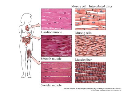
Vertebrates possess three primary types of muscle tissue: cardiac, smooth, and skeletal. Each type is specialized for distinct physiological functions and is characterized by unique structural and regulatory features.
Cardiac Muscle: Found only in the heart, responsible for pumping blood. Cells are branched, striated, and interconnected by gap junctions.
Smooth Muscle: Located in walls of internal organs (e.g., digestive tract, blood vessels). Cells are spindle-shaped, non-striated, and contract involuntarily.
Skeletal Muscle: Attached to bones, responsible for voluntary movements. Cells are long, multinucleate, and striated.
Structural Comparison
All muscle types utilize actin and myosin filaments for contraction, but their arrangement and control mechanisms differ.
Cardiac and Skeletal Muscle: Striated due to regular arrangement of actin and myosin.
Smooth Muscle: Non-striated; actin and myosin are irregularly arranged.
Skeletal Muscle Structure
Muscle Fiber Organization
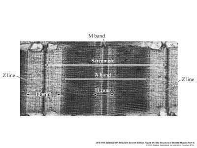
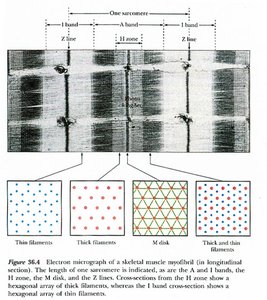
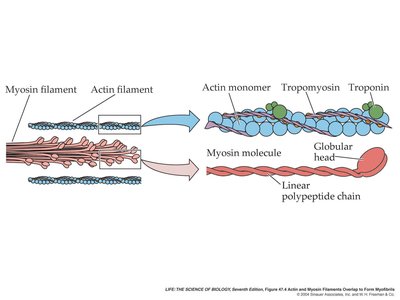
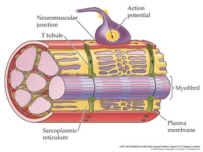
Skeletal muscle fibers are large, multinucleate cells packed with myofibrils. Each myofibril consists of repeating units called sarcomeres, the fundamental contractile unit.
Muscle fibers: Single cells containing many nuclei.
Myofibrils: Bundles of actin (thin) and myosin (thick) filaments.
Sarcomere: Bounded by Z lines, contains A band (myosin), I band (actin), H zone (non-overlapping region), and M band (myosin support proteins).
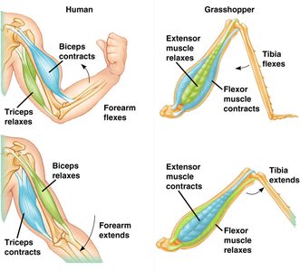
Antagonistic Muscle Pairs
Movement around joints is achieved by antagonistic pairs of muscles: one contracts while the other relaxes. Ligaments hold bones together, and tendons attach muscles to bones.
Sarcomere Structure and Function
Detailed Sarcomere Anatomy
The sarcomere is the basic unit of muscle contraction. Its structure is defined by the arrangement of actin and myosin filaments, supported by proteins such as titin.
Z line: Anchors actin filaments.
A band: Contains all myosin filaments.
M band: Supports myosin filaments.
H zone and I band: Regions where actin and myosin do not overlap in relaxed muscle.
Titin: Runs from Z line to Z line, stabilizing myosin bundles.
Muscle Contraction Mechanism
Sliding Filament Theory
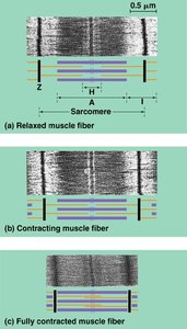
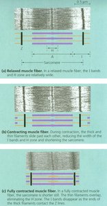
Muscle contraction occurs as actin and myosin filaments slide past each other, shortening the sarcomere. This process is known as the sliding filament theory.
During contraction, the H zone and I band narrow, and Z lines move toward the A band.
Myosin heads bind to actin, forming cross-bridges and pulling actin filaments inward.
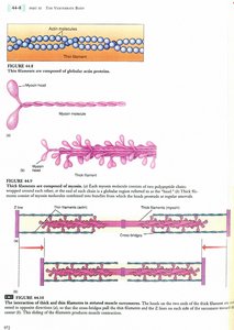

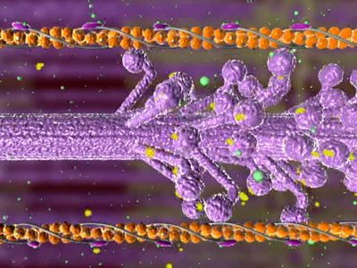
Actin and Myosin Structure
Actin filaments are composed of two chains of actin molecules twisted together, associated with regulatory proteins troponin and tropomyosin. Myosin filaments consist of many myosin molecules, each with a globular head capable of binding actin and hydrolyzing ATP.
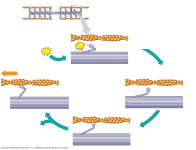
The Contractile Cycle
The contractile cycle involves several steps:
Myosin head binds ATP and is in a low-energy state.
ATP is hydrolyzed to ADP and Pi, energizing the myosin head.
Myosin head binds actin, forming a cross-bridge.
Release of ADP and Pi triggers the power stroke, sliding actin past myosin.
Binding of new ATP releases myosin from actin, restarting the cycle.
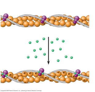
Regulation by Calcium, Troponin, and Tropomyosin
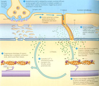
Muscle contraction is regulated by the presence of Ca2+ ions. At rest, troponin and tropomyosin block myosin binding sites on actin. When Ca2+ binds to troponin, the complex changes shape, exposing binding sites and allowing contraction.
Neural Control and Excitation-Contraction Coupling
Motor Neurons and Action Potentials
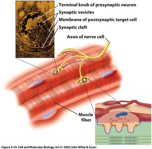
Skeletal muscle contraction is initiated by action potentials from motor neurons. Each motor neuron can stimulate multiple muscle fibers, forming a motor unit. Depolarization opens sodium channels, generating action potentials in the muscle fiber.
T-tubules and Sarcoplasmic Reticulum
Action potentials travel deep into the muscle fiber via T-tubules, which are continuous with the plasma membrane. T-tubules are closely associated with the sarcoplasmic reticulum (SR), a specialized organelle that stores Ca2+. Upon stimulation, Ca2+ is released from the SR into the cytoplasm, triggering contraction.
Muscle Fiber Types and Athletic Performance
Types of Skeletal Muscle Fibers
Skeletal muscle fibers are classified based on their metabolic properties and contraction speed:
Type I (Slow Oxidative, Red): High myoglobin, many mitochondria, fatigue-resistant, optimal for endurance activities.
Type IIa (Fast Oxidative, Red): High myoglobin, rapid ATP splitting, resistant to fatigue, suited for middle-distance activities.
Type IIb (Fast Glycolytic, White): Low myoglobin, few mitochondria, fatigues easily, optimal for short, intense activities.
Fiber Type | Myoglobin | Mitochondria | Fatigue Resistance | Optimal Activity |
|---|---|---|---|---|
Type I (Slow Oxidative) | High | Many | High | Endurance (e.g., marathon) |
Type IIa (Fast Oxidative) | High | Many | Moderate | Middle distance |
Type IIb (Fast Glycolytic) | Low | Few | Low | Sprinting, weightlifting |
Exercise and Muscle Adaptation
Exercise influences muscle fiber composition and function:
Strength training: Increases actin and myosin filaments, enlarging muscles.
Aerobic exercise: Enhances oxidative capacity, increases capillary density, myoglobin content, and mitochondria.
Drugs Affecting Muscle Function
Curare and Neuromuscular Blockade
Curare is a plant-derived toxin that blocks acetylcholine (ACh) receptors at the neuromuscular junction, preventing muscle contraction and causing paralysis.
Botulinum Toxin (Botox)
Botulinum toxin, produced by Clostridium botulinum, inhibits ACh release, causing paralysis. Used therapeutically for muscle spasms and cosmetically for wrinkle reduction. Effects are temporary and localized, but improper administration can cause serious side effects.
Summary Table: Key Muscle Components
Component | Function |
|---|---|
Tropomyosin | Blocks myosin-binding sites on actin |
Sarcoplasmic reticulum | Stores calcium ions in muscle |
Troponin | Calcium binding protein involved in muscle contraction |
T-tubule | Propagates action potential into the middle of a myofiber |
Thick filament | Responsible for higher density of A-band in striated muscle |
Acetylcholine | Initiates depolarization of a myofiber |
Practice Questions
Multiple Choice Example
Which of the following is the correct sequence that describes the excitation and contraction of a skeletal muscle fiber?
An action potential in a motor neuron causes the axon to release acetylcholine, which depolarizes the muscle cell membrane.
Transverse (T) tubules transmit the depolarization to the sarcoplasmic reticulum.
Calcium is released into the cytoplasm.
Tropomyosin is shifted, which unblocks the cross-bridge binding sites.
The thin filaments are ratcheted across the thick filaments by the heads of the myosin molecules using energy from ATP.
Correct answer: E) 5 → 3 → 2 → 1 → 4
Matching Example
Function | Component |
|---|---|
Blocks myosin-binding sites on actin | Tropomyosin |
Stores calcium ions in muscle | Sarcoplasmic reticulum |
Calcium binding protein involved in muscle contraction | Troponin |
Propagates action potential into the middle of a myofiber | T-tubule |
Responsible for higher density of A-band in striated muscle | Thick filament |
Initiates depolarization of a myofiber | Acetylcholine |
Key Equations
ATP Hydrolysis in Muscle Contraction
The hydrolysis of ATP by myosin provides energy for muscle contraction:
Conclusion
Muscle structure and function are central to vertebrate physiology, enabling movement, circulation, and organ function. Understanding the molecular mechanisms of contraction, regulation, and adaptation is essential for biology students.
