 Back
BackMuscle Structure and Function in Vertebrates
Study Guide - Smart Notes
 Tailored notes based on your materials, expanded with key definitions, examples, and context.
Muscle Structure and Function in Vertebrates
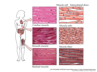
Types of Muscle Tissue
Vertebrates possess three main types of muscle tissue, each with distinct structures and functions. All muscle types utilize actin and myosin for contraction, but their organization and control mechanisms differ.
Cardiac Muscle: Found only in the heart, cardiac muscle cells are branched, striated, and interconnected by intercalated discs, which facilitate rapid electrical communication. Cardiac muscle contracts involuntarily and can generate its own rhythmic action potentials due to pacemaker cells.
Smooth Muscle: These spindle-shaped cells have a single nucleus and lack striations due to irregular arrangement of actin and myosin. Smooth muscle is found in the walls of hollow organs (e.g., digestive tract, blood vessels) and contracts involuntarily to move substances through the body.
Skeletal Muscle: Responsible for voluntary movements, skeletal muscle fibers are long, multinucleate, and striated due to the regular arrangement of actin and myosin. These muscles attach to bones via tendons and enable movement by contracting in antagonistic pairs.
Organization of Skeletal Muscle
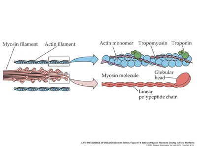
Skeletal muscle is composed of bundles of muscle fibers, each containing numerous myofibrils. Myofibrils are made up of repeating units called sarcomeres, the fundamental contractile units of muscle.
Muscle Fiber: A single, multinucleate cell packed with myofibrils.
Myofibril: A chain of sarcomeres aligned end-to-end.
Sarcomere: Bounded by Z lines, containing thick (myosin) and thin (actin) filaments. The arrangement of these filaments creates the striated appearance of skeletal muscle.
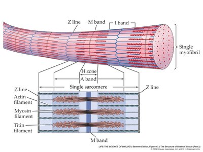
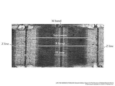
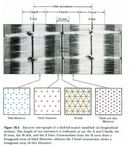
Sarcomere Structure and Sliding Filament Theory
The sarcomere contains several distinct regions:
Z line: Anchors thin filaments (actin).
A band: Region containing the entire length of thick filaments (myosin).
I band: Region with only thin filaments.
H zone: Central region with only thick filaments.
M band: Supports the arrangement of myosin filaments.
During contraction, actin and myosin filaments slide past each other, shortening the sarcomere without changing the length of the filaments themselves. This is known as the sliding filament theory.
Molecular Mechanism of Muscle Contraction
Muscle contraction is powered by the interaction of actin and myosin, regulated by ATP and calcium ions (Ca2+).
Myosin: Thick filament protein with heads that bind to actin and hydrolyze ATP.
Actin: Thin filament protein forming a double helix, associated with regulatory proteins troponin and tropomyosin.
Troponin and Tropomyosin: Block myosin-binding sites on actin in resting muscle. Ca2+ binding to troponin causes a conformational change, exposing the binding sites and allowing contraction.

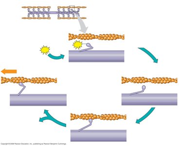
The Contractile Cycle
Myosin head binds ATP and is in a low-energy state.
ATP hydrolysis "cocks" the myosin head into a high-energy conformation.
Myosin binds to actin, forming a cross-bridge.
Release of ADP and Pi triggers the power stroke, sliding actin past myosin.
Binding of new ATP releases myosin from actin, restarting the cycle.
Role of Calcium in Muscle Contraction
Calcium ions are released from the sarcoplasmic reticulum in response to an action potential. Ca2+ binds to troponin, causing tropomyosin to shift and expose myosin-binding sites on actin, enabling contraction.

Neural Control of Muscle Contraction
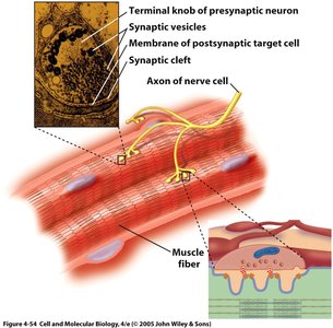
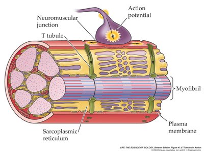
Skeletal muscle contraction is initiated by action potentials from motor neurons. Each motor neuron and the muscle fibers it innervates form a motor unit. The neuromuscular junction is the synapse where the neuron communicates with the muscle fiber via the neurotransmitter acetylcholine (ACh).
Action potential in the neuron triggers ACh release.
ACh binds to receptors on the muscle fiber, depolarizing the membrane and generating an action potential in the muscle.
The action potential travels along the sarcolemma and into the muscle fiber via T-tubules, triggering Ca2+ release from the sarcoplasmic reticulum.
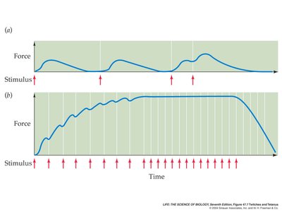
Summation and Tetanus
Muscle fibers can be stimulated repeatedly before they fully relax, leading to increased force production (summation). At high stimulation frequencies, maximal tension is achieved, known as tetanus.
Temporal Summation: Increased frequency of stimulation increases force.
Spatial Summation: Recruitment of more motor units increases force.
Types of Skeletal Muscle Fibers
Skeletal muscle fibers are classified based on their metabolic properties and contraction speed:
Type I (Slow Oxidative, Red Fibers): High myoglobin, many mitochondria, fatigue-resistant, optimal for endurance activities.
Type IIa (Fast Oxidative, Red Fibers): Intermediate properties, resistant to fatigue, suited for middle-distance activities.
Type IIb (Fast Glycolytic, White Fibers): Low myoglobin, few mitochondria, fatigue quickly, optimal for short, intense activities.
The proportion of fiber types is influenced by genetics and training. Aerobic exercise increases oxidative capacity, while strength training increases muscle size.
Muscle Paralysis and Drugs
Certain toxins and drugs can interfere with neuromuscular transmission, leading to paralysis:
Curare: Blocks ACh receptors, preventing muscle contraction and causing paralysis.
Botulinum Toxin (Botox): Inhibits ACh release at the neuromuscular junction, causing muscle paralysis. Used medically for various conditions and cosmetically to reduce wrinkles.
Clinical and Historical Context
Foodborne botulism, caused by Clostridium botulinum contamination in improperly sterilized foods, led to significant public health reforms in the early 20th century. Modern uses of botulinum toxin include treatment for muscle spasms, migraines, and other disorders, but improper administration can cause serious side effects.
Sample Exam Questions
Sequence of Skeletal Muscle Contraction: Action potential in motor neuron → ACh release → Muscle cell depolarization → T-tubule transmission → Ca2+ release → Tropomyosin shift → Cross-bridge cycling.
Motor Unit: One motor neuron and all the muscle fibers it innervates.
Matching Terms:
Blocks myosin-binding sites on actin: Tropomyosin
Stores calcium ions in muscle: Sarcoplasmic reticulum
Calcium binding protein: Troponin
Propagates action potential: T-tubule
Responsible for A-band density: Thick filament
Initiates depolarization: Acetylcholine
Additional info: This guide integrates textbook-level explanations and diagrams to provide a comprehensive overview of muscle structure and function, suitable for college-level biology students.
