 Back
BackMuscle Structure and Function: Mechanisms of Animal Movement
Study Guide - Smart Notes
 Tailored notes based on your materials, expanded with key definitions, examples, and context.
Muscle Structure and Function
Types and Functions of Muscles
Muscle tissue is essential for movement in animals and is classified into three main types, each with distinct structures and functions:
Skeletal Muscle: Voluntary muscle attached to bones, responsible for body movement and posture. It is striated and multinucleated.
Cardiac Muscle: Involuntary muscle found only in the heart, characterized by striations and intercalated discs for synchronized contraction.
Smooth Muscle: Involuntary, non-striated muscle found in the walls of internal organs (e.g., intestines, blood vessels), responsible for slow, sustained contractions.
Function: Muscles convert chemical energy (ATP) into mechanical work, enabling movement, maintaining posture, and regulating internal processes.
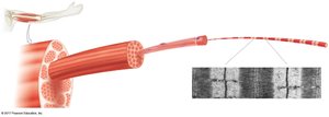
Structure of Skeletal Muscle
Skeletal muscle is organized into bundles of muscle fibers, each containing myofibrils composed of repeating units called sarcomeres. Sarcomeres are the fundamental contractile units of muscle, defined by the arrangement of actin (thin) and myosin (thick) filaments.
Muscle Fiber: A single muscle cell, multinucleated, containing many myofibrils.
Myofibril: Cylindrical structures within muscle fibers, made up of sarcomeres in series.
Sarcomere: The basic contractile unit, bordered by Z-discs, containing overlapping actin and myosin filaments.
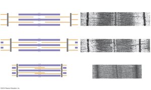
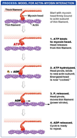
Sliding Filament Theory
The sliding filament theory explains how muscles contract at the molecular level. During contraction, myosin heads bind to actin filaments and pull them toward the center of the sarcomere, shortening the muscle without changing the length of the filaments themselves.
Key Steps:
Myosin heads attach to actin, forming cross-bridges.
Using energy from ATP hydrolysis, myosin heads pivot, pulling actin filaments inward (power stroke).
ATP binds to myosin, causing it to detach from actin and reset for another cycle.
Result: Sarcomeres shorten, leading to muscle contraction.
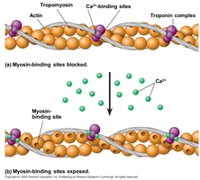
Regulation of Muscle Contraction
Muscle contraction is regulated by the availability of calcium ions (Ca2+) and the proteins troponin and tropomyosin. In a relaxed muscle, tropomyosin blocks myosin-binding sites on actin. When Ca2+ is released from the sarcoplasmic reticulum, it binds to troponin, causing a conformational change that moves tropomyosin and exposes the binding sites, allowing contraction to occur.
Troponin: A regulatory protein that binds Ca2+ and controls the position of tropomyosin.
Tropomyosin: A protein that blocks myosin-binding sites on actin in the absence of Ca2+.
Calcium Ions: Trigger the exposure of binding sites, initiating contraction.
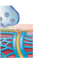
Motor Synapses (Neuromuscular Junction)
The neuromuscular junction is the synapse between a motor neuron and a skeletal muscle fiber. When an action potential reaches the axon terminal, it triggers the release of the neurotransmitter acetylcholine (ACh), which binds to receptors on the muscle cell membrane, initiating a muscle action potential and subsequent contraction.
Motor Neuron: Transmits the nerve impulse to the muscle fiber.
Acetylcholine (ACh): Neurotransmitter that stimulates muscle contraction.
Muscle Action Potential: Electrical signal that triggers Ca2+ release and contraction.
Muscle Twitches and Cramps
A muscle twitch is a single, brief contraction and relaxation cycle in a muscle fiber, typically resulting from a single action potential. Muscle cramps are involuntary, sustained contractions often caused by electrolyte imbalances, dehydration, or overuse.
Twitch: The response of a muscle to a single stimulus.
Cramps: Prolonged, involuntary contractions that can be painful and are often due to physiological disturbances.
Muscle Growth
Muscle growth, or hypertrophy, occurs when muscle fibers increase in size due to repeated stimulation, such as resistance training. This process involves the synthesis of new contractile proteins and the addition of myofibrils within muscle fibers.
Hypertrophy: Increase in muscle fiber size due to increased protein synthesis.
Stimuli: Resistance exercise, hormones (e.g., growth hormone, testosterone), and adequate nutrition.
