 Back
BackMuscle Structure, Function, and Regulation in Animals
Study Guide - Smart Notes
 Tailored notes based on your materials, expanded with key definitions, examples, and context.
Muscle Structure and Function
Overview of Muscle Types
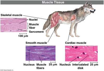
Muscle tissue is essential for movement, support, and various physiological functions in animals. There are three main types of muscle: skeletal, cardiac, and smooth, each with distinct structural and functional properties.
Skeletal muscle: Attaches to bones, is striated, and is under voluntary control.
Cardiac muscle: Found in the heart, striated, involuntary, and cells are connected by intercalated discs.
Smooth muscle: Located in walls of hollow organs, not striated, involuntary, and controlled by the autonomic nervous system.
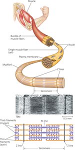
Skeletal Muscle Structure
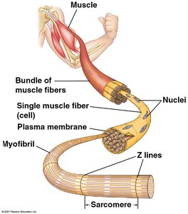
Skeletal muscle is composed of bundles of long, multinucleated muscle fibers. Each fiber contains several myofibrils, which are made up of repeating units called sarcomeres. Sarcomeres are the fundamental contractile units of muscle.
Myofibril: A chain of sarcomeres within a muscle fiber.
Sarcomere: Contains thin (actin) and thick (myosin) filaments, bordered by Z-lines.
Striated appearance: Due to the alignment of sarcomeres in adjacent myofibrils.
The Sliding Filament Model of Muscle Contraction
Muscle contraction occurs when thin and thick filaments slide past each other, powered by ATP-driven myosin activity. The filaments themselves do not change length; instead, their overlap increases, shortening the sarcomere and thus the muscle.
Thin filaments: Composed of actin, anchored at Z-lines.
Thick filaments: Composed of myosin, anchored at the M-line.
Contraction: Myosin heads bind to actin, pull the filaments, and release, repeating the cycle.
Extension: Passive process; muscle returns to resting length.
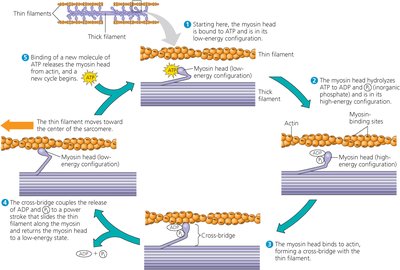
Mechanism of the Sliding Filament Cycle
The contraction cycle involves several steps, each powered by ATP:
Myosin head binds ATP and detaches from actin.
ATP hydrolysis energizes the myosin head.
Myosin binds to actin, forming a cross-bridge.
Power stroke: Myosin releases ADP and Pi, pulling actin toward the center.
New ATP binds, releasing myosin from actin, and the cycle repeats.
Regulation of Muscle Contraction
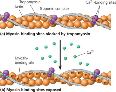
Role of Calcium and Regulatory Proteins
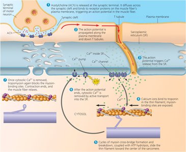
Muscle contraction is tightly regulated by calcium ions (Ca2+) and regulatory proteins. At rest, tropomyosin blocks myosin-binding sites on actin. Upon stimulation, Ca2+ binds to the troponin complex, shifting tropomyosin and exposing binding sites.
Tropomyosin: Covers myosin-binding sites on actin.
Troponin complex: Binds Ca2+ and moves tropomyosin.
Ca2+ release: Triggered by motor neuron stimulation.
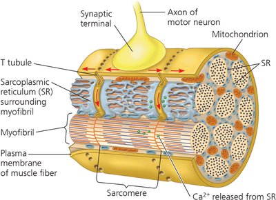
Neural Control of Muscle Contraction
Motor neurons release acetylcholine at the neuromuscular junction, generating action potentials in the muscle fiber. These action potentials travel along T-tubules, causing Ca2+ release from the sarcoplasmic reticulum and initiating contraction.
Motor neuron: Releases acetylcholine, stimulating muscle fiber.
T-tubules: Invaginations of the plasma membrane that transmit action potentials.
Sarcoplasmic reticulum: Stores and releases Ca2+.
Motor Units and Muscle Contractility
A motor unit consists of a single motor neuron and all the muscle fibers it controls. The strength of muscle contraction depends on the number of motor units activated and the frequency of stimulation.
All-or-none twitch: Individual muscle fibers contract fully or not at all.
Graded contraction: Whole muscle contraction varies in strength.
Recruitment: Activation of additional motor units increases force.
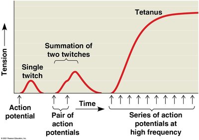
Summation, Tetanus, and Fatigue
Muscle tension can be modulated by the rate of stimulation. If action potentials arrive before relaxation, their effects sum, increasing tension. High-frequency stimulation leads to tetanus, a sustained contraction. Prolonged contraction causes fatigue due to ATP depletion and ion imbalance.
Summation: Addition of twitches increases tension.
Tetanus: Maximum, sustained contraction.
Fatigue: Loss of contractile ability due to resource depletion.
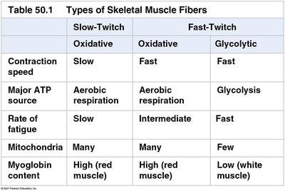
Types of Skeletal Muscle Fibers
Classification of Muscle Fibers
Skeletal muscle fibers are classified based on contraction speed and metabolic properties. Slow-twitch fibers are adapted for endurance, while fast-twitch fibers are suited for rapid, powerful contractions.
Slow-twitch (oxidative): Slow contraction, high endurance, many mitochondria, high myoglobin.
Fast-twitch (oxidative): Fast contraction, intermediate endurance, many mitochondria, high myoglobin.
Fast-twitch (glycolytic): Fast contraction, low endurance, few mitochondria, low myoglobin.
Type | Contraction Speed | Major ATP Source | Rate of Fatigue | Mitochondria | Myoglobin Content |
|---|---|---|---|---|---|
Slow-Twitch (Oxidative) | Slow | Aerobic respiration | Slow | Many | High (red muscle) |
Fast-Twitch (Oxidative) | Fast | Aerobic respiration | Intermediate | Many | High (red muscle) |
Fast-Twitch (Glycolytic) | Fast | Glycolysis | Fast | Few | Low (white muscle) |
Muscle Tissue Types
Skeletal, Cardiac, and Smooth Muscle
Each muscle type has unique structural and functional characteristics:
Skeletal muscle: Striated, voluntary, multinucleated fibers.
Cardiac muscle: Striated, involuntary, intercalated discs for electrical connectivity.
Smooth muscle: Non-striated, involuntary, spindle-shaped cells.
Muscle Contraction and Movement
Role of Skeletons in Movement
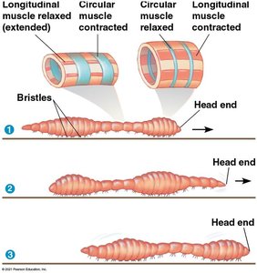
Muscle contraction produces movement by acting on skeletal elements. Skeletons provide support, protection, and leverage for muscle action. Two muscles attached to the same section of the skeleton often work antagonistically to move body parts back and forth.
Support: Skeletons maintain body shape and protect organs.
Movement: Muscles contract to move skeleton parts.
Antagonistic pairs: One muscle contracts while the other relaxes.
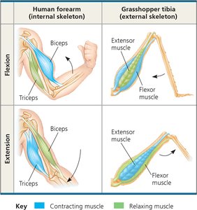
Types of Skeletal Systems
Animals possess different types of skeletal systems:
Hydrostatic skeleton: Fluid-filled compartments; movement by changing shape (e.g., worms).
Exoskeleton: Hard external shell; muscles attach to the shell (e.g., arthropods).
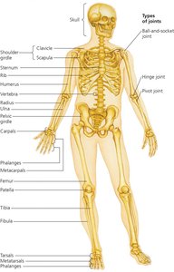
Endoskeleton: Internal skeleton of cartilage and bone (e.g., humans).
Tendons and Ligaments
Tendons attach muscles to bones, while ligaments hold bones together at joints. These connective tissues are essential for transmitting force and stabilizing joints during movement.
Tendons: Connect muscle to bone.
Ligaments: Connect bone to bone at joints.
Sensory Systems and Motor Output
Integration of Sensory Input and Motor Output
Sensory inputs are processed by the nervous system, resulting in motor outputs that drive muscle activity and behavior. The central nervous system integrates sensory information and coordinates appropriate responses.
Sensory input: Information from receptors (e.g., touch, vision).
Integration: Processing in the central nervous system.
Motor output: Activation of effectors (muscles, glands).
Summary of Key Concepts
Muscle contraction is powered by myosin and regulated by Ca2+ and regulatory proteins.
Motor units and neural stimulation control muscle contractility and tension.
Movement requires a skeleton, which can be hydrostatic, exoskeletal, or endoskeletal.
Skeletal muscle fibers are classified by contraction speed and metabolic properties.
Sensory systems and motor output are integrated by the nervous system to produce behavior.
