 Back
BackChapter 15
Study Guide - Smart Notes
 Tailored notes based on your materials, expanded with key definitions, examples, and context.
Mutation, DNA Repair, and Cancer
Mutation: Definition and Importance
Mutations are heritable changes in the genetic material and are essential for the continuity of life. They serve as the source of genetic variation, which is fundamental for natural selection. However, most new mutations are more likely to be harmful than beneficial. DNA repair systems exist to reverse DNA damage, and cancer is a disease caused by gene mutations.
Types of Gene Mutations and Their Effects
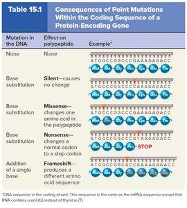
Gene mutations can affect the amino acid sequence of proteins, leading to various consequences:
Silent Mutation: Does not alter the amino acid sequence due to the degeneracy of the genetic code.
Missense Mutation: Changes a single amino acid in a polypeptide. The effect depends on the similarity of the substituted amino acid to the original. Example: Sickle-cell disease.
Nonsense Mutation: Changes a normal codon to a stop codon, producing a truncated polypeptide.
Frameshift Mutation: Addition or deletion of nucleotides (not multiples of 3), resulting in a completely different amino acid sequence downstream from the mutation.
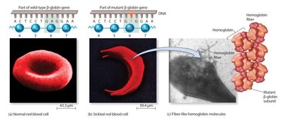
Example: Sickle-cell Disease
Sickle-cell disease is caused by a missense mutation in the β-globin gene, resulting in abnormal hemoglobin and sickled red blood cells.
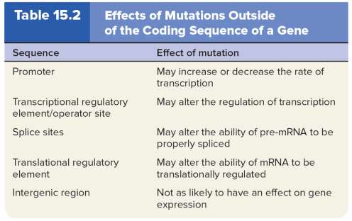
Mutations Outside Coding Sequences
Mutations can also occur outside coding regions, affecting gene regulation and expression:
Promoter: May increase or decrease the rate of transcription.
Transcriptional regulatory element/operator site: May alter regulation of transcription.
Splice sites: May affect the ability of pre-mRNA to be properly spliced.
Translational regulatory element: May affect the ability of mRNA to be translationally regulated.
Intergenic region: Less likely to affect gene expression.
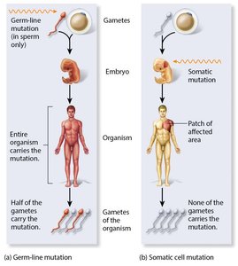
Germ-line vs. Somatic Cell Mutations
The time and location of a mutation determine its severity and heritability:
Germ-line mutations: Occur in gametes or their progenitor cells and are heritable.
Somatic mutations: Occur in other body cells and can result in genetic mosaics, but are not heritable.
Causes of Gene Mutations
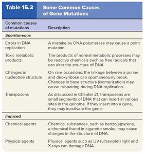
Mutations can be spontaneous or induced:
Spontaneous mutations: Result from errors in DNA replication, toxic metabolic products, changes in nucleotide structure, or transposons.
Induced mutations: Caused by environmental agents (mutagens) such as chemicals or physical agents.
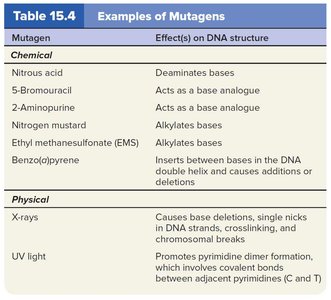
Mutagens and Their Effects
Mutagens are agents that alter DNA, leading to mutations:
Chemical mutagens: Modify nucleotide structure, act as base analogues, or insert between bases.
Physical mutagens: Include ionizing radiation (X-rays, gamma rays) and nonionizing radiation (UV light).
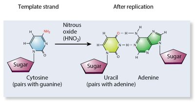
Nitrous Oxide as a Mutagen
Nitrous oxide deaminates cytosine, converting it to uracil, which pairs with adenine instead of guanine, leading to mutations.
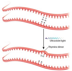
UV Light and Thymine Dimers
UV light causes the formation of thymine dimers, which can disrupt DNA replication and lead to mutations.
DNA Repair Mechanisms
Organisms have evolved mechanisms to detect and repair DNA damage:
Direct repair: Enzyme recognizes and corrects incorrect DNA structure.
Nucleotide excision repair (NER): Removes and replaces a damaged DNA region using the undamaged strand as a template.
Methyl-directed mismatch repair: Detects base pair mismatches and removes the surrounding DNA strand for replacement.
NER and Human Genetic Disease
Defects in NER are associated with genetic diseases such as Xeroderma pigmentosum, Cockayne’s syndrome, and PIBIDS, all characterized by photosensitivity due to inability to repair UV-induced lesions.
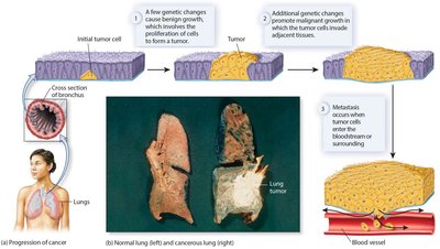
Cancer: General Information
Cancer is a disease of multicellular organisms characterized by uncontrolled cell division. It can originate from a single cell and progress from benign to malignant, invasive, and metastatic stages. Most cancers are not inherited, but exposure to carcinogens (mutagens) increases risk.
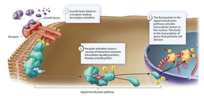
Oncogenes and Cell Division
Cell division is regulated by growth factors. Mutations in genes for cell growth signaling proteins can convert them into oncogenes, which promote cancer by keeping the cell division pathway permanently active.
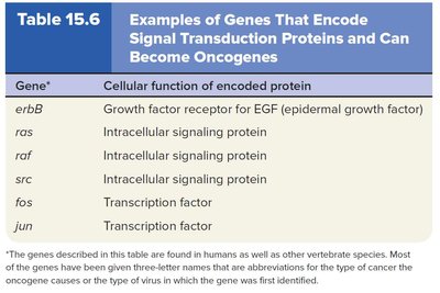
Examples of Oncogenes
erbB: Growth factor receptor for EGF
ras: Intracellular signaling protein
raf, src: Intracellular signaling proteins
fos, jun: Transcription factors
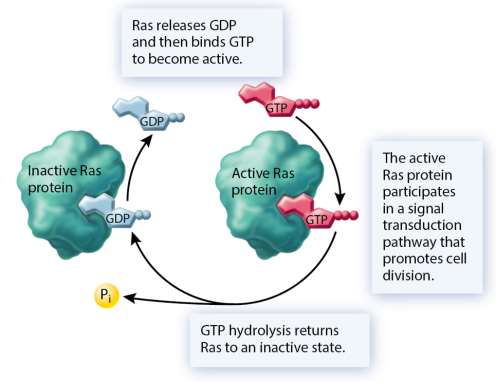
Ras Protein and Cancer
Ras is an intracellular signaling protein that hydrolyzes GTP. Oncogenic mutations may decrease Ras's ability to hydrolyze GTP or increase GDP/GTP exchange, keeping the signaling pathway constantly active.
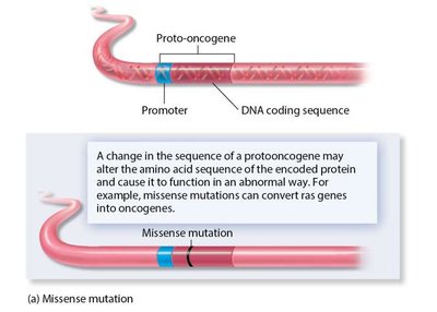
Proto-oncogenes and Genetic Changes
Proto-oncogenes are normal genes that can become oncogenes through:
Missense mutations: Change amino acid sequence, potentially converting proto-oncogenes to oncogenes.
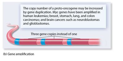
Gene amplifications: Increase copy number, resulting in excess protein.
Chromosomal translocations: Create chimeric genes associated with certain tumors.
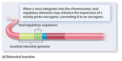
Retroviral insertions: Viral DNA inserts near proto-oncogenes, enhancing expression.
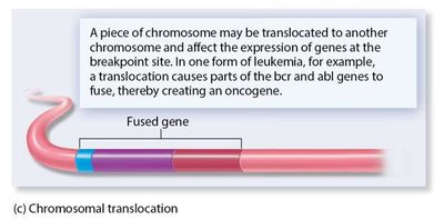
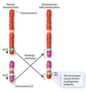
Chromosomal Translocation Example
Translocation between chromosomes 9 and 22 creates a fused gene (bcr-abl), causing chronic myelogenous leukemia.
Viruses That Cause Cancer
Virus | Description |
|---|---|
Rous sarcoma virus | Causes sarcomas in chickens |
Simian sarcoma virus | Causes sarcomas in monkeys |
Abelson leukemia virus | Causes leukemia in mice |
Hardy-Zuckerman 4 feline sarcoma virus | Causes sarcomas in cats |
Hepatitis B | Causes liver cancer in several species, including humans |
Tumor-Suppressor Genes
Tumor-suppressor genes prevent cancerous growth by maintaining genome integrity and inhibiting cell division. Checkpoint proteins, such as p53, halt cell cycle progression if DNA damage is detected.
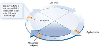
p53 Protein
Acts at the G1 checkpoint to prevent progression to S phase if DNA is damaged.
If DNA cannot be repaired, p53 activates genes promoting apoptosis (programmed cell death).
Rb Protein and Retinoblastoma
Rb protein inhibits the transcription factor E2F, preventing cell division. Loss of both Rb gene copies results in uncontrolled cell division and retinoblastoma.
Loss of Tumor-Suppressor Gene Function
Mutation within the gene inactivates its function.
Chromosome loss removes tumor-suppressor genes.
Abnormal methylation of CpG islands near promoter regions silences gene expression.
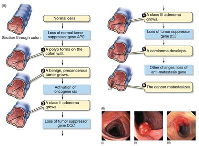
Cancer as a Series of Genetic Changes
Cancer typically requires multiple genetic changes, beginning with benign alterations and progressing to malignancy. Additional mutations can make cancer more difficult to treat.
Summary Table: Consequences of Point Mutations
Mutation in the DNA | Effect on polypeptide | Example |
|---|---|---|
None | No change | Normal sequence |
Base substitution | Silent—causes no change | Same amino acid sequence |
Base substitution | Missense—changes one amino acid | Sickle-cell disease |
Base substitution | Nonsense—changes normal codon to stop codon | Truncated polypeptide |
Addition or deletion | Frameshift—produces different amino acid sequence | Altered sequence downstream |
Summary Table: Effects of Mutations Outside Coding Sequence
Sequence | Effect of mutation |
|---|---|
Promoter | May increase or decrease transcription rate |
Transcriptional regulatory element/operator site | May alter regulation of transcription |
Splice sites | May affect pre-mRNA splicing |
Translational regulatory element | May affect mRNA translation |
Intergenic region | Less likely to affect gene expression |
Summary Table: Common Causes of Gene Mutations
Common causes | Description |
|---|---|
Errors in DNA replication | DNA polymerase mistakes |
Toxic metabolic products | Reactive chemicals from metabolism |
Changes in nucleotide structure | Spontaneous base linkage breakage |
Transposons | Mobile DNA segments |
Chemical agents | Mutagens like benzopyrene |
Physical agents | UV light, X-rays |
Summary Table: Examples of Mutagens
Mutagen | Effect(s) on DNA structure |
|---|---|
Nitrous acid | Deaminates bases |
5-Bromouracil | Acts as base analogue |
2-Aminopurine | Acts as base analogue |
Nitrogen mustard | Alkylates bases |
Ethyl methanesulfonate (EMS) | Alkylates bases |
Benzo(a)pyrene | Inserts between bases, causes additions/deletions |
X-rays | Causes base deletions, nicks, crosslinking, breaks |
UV light | Promotes pyrimidine dimer formation |
Summary Table: Genes That Encode Signal Transduction Proteins
Gene | Cellular function |
|---|---|
erbB | Growth factor receptor for EGF |
ras | Intracellular signaling protein |
raf | Intracellular signaling protein |
src | Intracellular signaling protein |
fos | Transcription factor |
jun | Transcription factor |
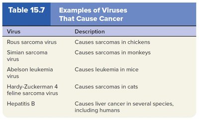
Summary Table: Examples of Viruses That Cause Cancer
Virus | Description |
|---|---|
Rous sarcoma virus | Causes sarcomas in chickens |
Simian sarcoma virus | Causes sarcomas in monkeys |
Abelson leukemia virus | Causes leukemia in mice |
Hardy-Zuckerman 4 feline sarcoma virus | Causes sarcomas in cats |
Hepatitis B | Causes liver cancer in several species, including humans |
Summary Table: Cell Cycle Checkpoints
Checkpoint | Function |
|---|---|
G1 checkpoint | Halts cell division if DNA damage is detected |
G2 checkpoint | Ensures DNA is fully replicated and undamaged |
Metaphase checkpoint | Ensures chromosomes are properly aligned |
Summary Table: Genetic Changes in Cancer Progression
Stage | Genetic change |
|---|---|
Benign | Initial genetic alteration |
Malignant | Additional mutations |
Metastatic | Further genetic changes, invasion, migration |
