 Back
BackMutations: Types, Mechanisms, and Biological Consequences
Study Guide - Smart Notes
 Tailored notes based on your materials, expanded with key definitions, examples, and context.
Mutations and Genetic Variation
Definition and Importance of Mutations
Mutations are changes in the genetic material (DNA sequence) of an organism. They are the primary source of genetic diversity in living cells and viruses, providing the raw material for evolution and adaptation. Mutations can result in new genes and, consequently, new proteins with altered functions.
Genetic diversity: Mutations introduce variation within populations, which is essential for natural selection.
Hereditary diseases: Some mutations can have adverse effects, leading to genetic disorders.
Types of Mutations
Large-Scale (Chromosomal) Mutations
Large-scale mutations involve chromosomal rearrangements that affect extensive segments of DNA. These can include deletions, duplications, inversions, and translocations, often resulting in significant phenotypic effects.
Small-Scale Mutations
Small-scale mutations affect only a single or a few nucleotides. The most common type is the point mutation, which involves a change in a single base pair. Even a single nucleotide change can lead to the production of an abnormal protein, as seen in many genetic diseases.
Transmission and Effects of Mutations
Inheritance and Disease
Mutations can be passed to offspring if they occur in germ cells. If a mutation adversely affects the phenotype, it is termed a genetic disorder or hereditary disease.
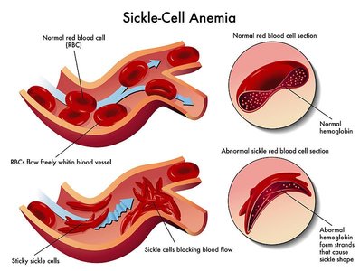
Case Study: Sickle-Cell Disease
Molecular Basis of Sickle-Cell Disease
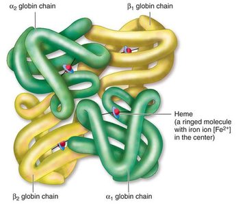
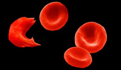
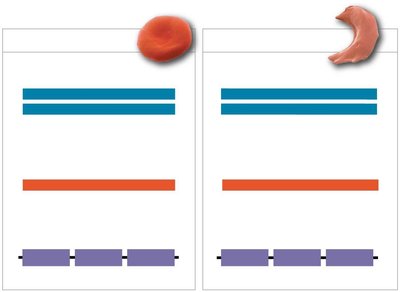
Sickle-cell disease is caused by a point mutation in the gene encoding the β-globin polypeptide of hemoglobin. This single nucleotide change leads to the production of an abnormal hemoglobin protein, which causes red blood cells to become sickle-shaped under low oxygen conditions.
Normal β-globin: Encodes glutamate (Glu)
Mutant β-globin: Encodes valine (Val) due to a single base substitution
Consequences at Multiple Levels
Protein level: The altered hemoglobin tends to clump together, distorting the red blood cell shape.
Cellular level: Sickle-shaped cells can block blood flow, leading to pain and fatigue.
Organism level: Reduced oxygen delivery and increased risk of complications.
Categories of Small-Scale Mutations
Point Mutations
Point mutations can be classified into:
Nucleotide-pair substitutions: Replacement of one nucleotide and its partner with another pair.
Nucleotide-pair insertions or deletions (Indels): Addition or loss of nucleotide pairs, which may cause frameshifts.
Types of Substitution Mutations
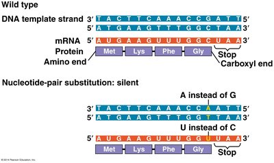
Silent mutations: No effect on the amino acid sequence due to redundancy in the genetic code.
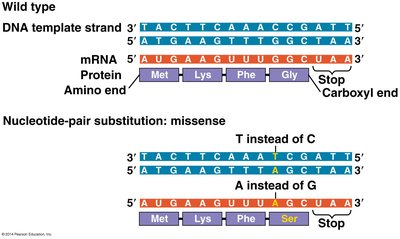
Missense mutations: Change one amino acid to another, potentially altering protein function.
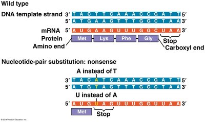
Nonsense mutations: Convert an amino acid codon into a stop codon, resulting in a truncated, usually nonfunctional protein.
Insertions and Deletions (Indels)
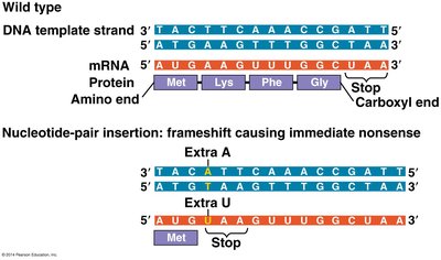
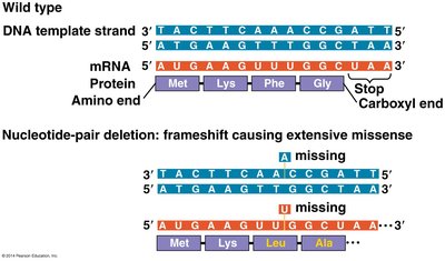
Insertions and deletions can have more severe effects than substitutions, especially if they alter the reading frame (frameshift mutations), which changes every amino acid downstream of the mutation.
Frameshift mutations: Caused by indels not in multiples of three, leading to extensive missense or premature stop codons.
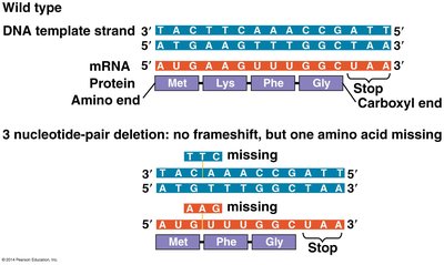
In-frame indels: Addition or deletion of three nucleotides removes or adds an amino acid without shifting the reading frame.
Genetic Code and Mutation Analysis
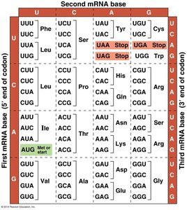
Using the Genetic Code Table
The genetic code table is essential for determining the effects of mutations on protein sequences. It allows translation of mRNA codons into amino acids.
Causes of DNA Mutations
Natural (Internal) Causes
Errors during DNA replication
Spontaneous chemical changes
Problems during cell division (e.g., recombination errors)
Induced (External) Causes
Chemical agents: Such as tobacco smoke and industrial chemicals
Physical agents: Non-ionizing radiation (e.g., UV light) and ionizing radiation (e.g., X-rays)
Viruses: Can integrate into the genome and disrupt gene function
DNA Proofreading and Repair Mechanisms
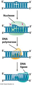
Nucleotide Excision Repair
Cells possess proofreading and repair systems to correct DNA damage. Nucleotide excision repair involves detection of damage, removal of the damaged section, synthesis of new DNA, and ligation of the backbone.
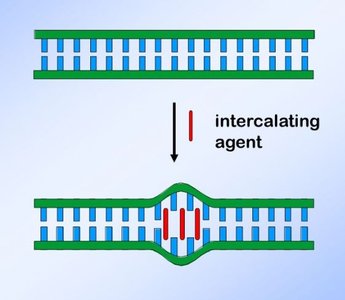
Intercalating Agents
Intercalating agents are flat, planar molecules that insert between DNA bases, distorting the DNA and interfering with replication and transcription.
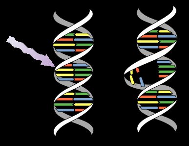
Pyrimidine Dimers and UV Damage
Non-ionizing radiation, such as UV light, can cause adjacent pyrimidines (usually thymines) to form covalent bonds, creating pyrimidine dimers that distort the DNA helix. These are typically repaired by nucleotide excision repair enzymes. Defects in these enzymes, as seen in Xeroderma pigmentosum, increase the risk of skin cancer.
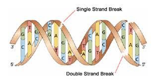
Ionizing Radiation
Ionizing radiation (e.g., X-rays) can cause single- and double-strand breaks in DNA, which are challenging for cells to repair and can lead to severe mutations or cell death.
Biological Consequences and Benefits of Mutations
Detrimental and Beneficial Effects
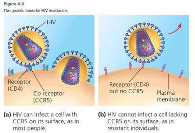
While many mutations are harmful, some can be beneficial or neutral. For example, the sickle-cell mutation provides resistance to malaria in endemic regions. Another example is the CCR5 Δ32 mutation, a 32-base-pair deletion in the CCR5 gene, which confers resistance to HIV infection by producing a nonfunctional receptor.
Additional info: Mutations are a driving force in evolution, and their effects depend on the context of the organism's environment and genetic background.
