 Back
BackNervous Systems: Structure, Function, and Disorders
Study Guide - Smart Notes
 Tailored notes based on your materials, expanded with key definitions, examples, and context.
Concept 1: Nervous Systems Consist of Circuits of Neurons and Supporting Cells
Overview of Nervous Systems
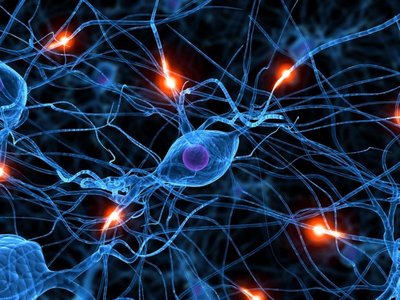
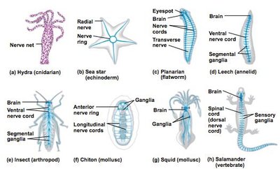
Nervous systems enable organisms to sense and respond to environmental changes. The complexity of nervous systems varies widely among animal groups, from simple nerve nets to highly organized brains and spinal cords.
Nerve Net: The simplest form of nervous system, found in cnidarians (e.g., Hydra), consists of interconnected neurons without a central brain.
Nerves: Bundles of axons that transmit signals in more complex animals.
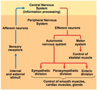
Central Nervous System (CNS): Composed of the brain and spinal cord; responsible for processing information.
Peripheral Nervous System (PNS): Consists of nerves and ganglia outside the CNS; transmits signals between the CNS and the rest of the body.
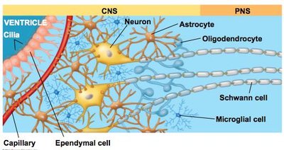
Glial Cells: Structure and Function
Glial cells, or glia, are non-neuronal cells that support and protect neurons in the nervous system.
Astrocytes: Nourish neurons, maintain the blood-brain barrier, and regulate the extracellular environment.
Oligodendrocytes (CNS) and Schwann Cells (PNS): Form myelin sheaths that insulate axons and speed up signal transmission.
Microglial Cells: Act as immune cells, removing debris and pathogens.
Ependymal Cells: Line the ventricles of the brain and help produce cerebrospinal fluid (CSF).
Organization of the Vertebrate Nervous System
The vertebrate nervous system is divided into the CNS and PNS, each with specialized structures and functions.
Brain: The main processing center for sensory information and responses.
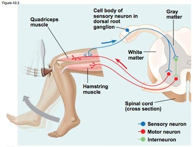
Spinal Cord: Conveys information to and from the brain and can produce reflexes independently.
Reflex: An automatic response to a stimulus, such as the knee-jerk reflex.
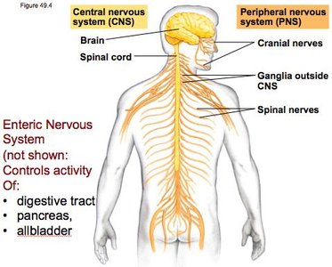
Peripheral Nervous System (PNS) Components
The PNS is divided into afferent (sensory) and efferent (motor) divisions. The efferent division includes the autonomic nervous system, which regulates involuntary functions.
Afferent Division: Transmits sensory information to the CNS.
Efferent Division: Carries commands from the CNS to muscles and glands.
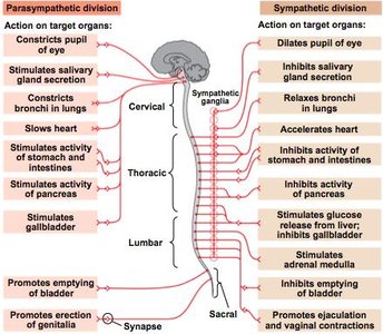
Autonomic Nervous System: Controls involuntary activities and is subdivided into:
Sympathetic Division (fight or flight)
Parasympathetic Division (rest and digest)
Enteric Division (regulates digestive tract)
Concept 2: The Vertebrate Brain is Regionally Specialized
Brain Regionalization and Evolution
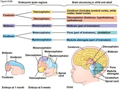
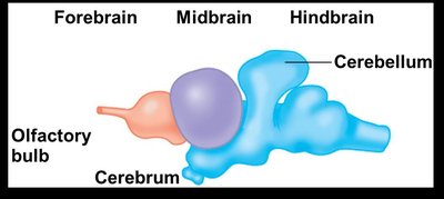
The vertebrate brain is divided into specialized regions, each responsible for distinct functions. Evolution has led to increased complexity and specialization in different vertebrate groups.
Forebrain: Involved in processing sensory information, reasoning, and voluntary movement.
Midbrain: Coordinates sensory input and motor output.
Hindbrain: Controls involuntary activities and coordinates movement.
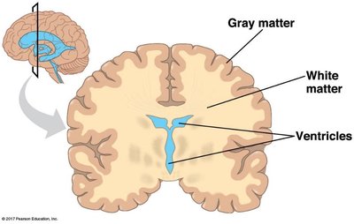
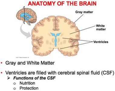
Gray Matter, White Matter, and Ventricles
The brain contains regions of gray matter (neuron cell bodies), white matter (myelinated axons), and fluid-filled ventricles.
Gray Matter: Contains neuron cell bodies and is involved in processing information.
White Matter: Composed of myelinated axons that transmit signals between brain regions.
Ventricles: Cavities filled with cerebrospinal fluid (CSF) that provide nutrition and protection.
Arousal, Sleep, and Biological Clocks
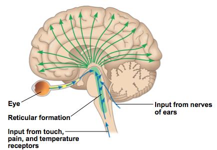
The brainstem and cerebrum regulate arousal and sleep. The reticular formation acts as a sensory filter, and the pineal gland releases melatonin to regulate sleep cycles. Circadian rhythms are controlled by the suprachiasmatic nucleus (SCN) in the hypothalamus.
Emotions and the Limbic System
The limbic system, including the amygdala, hippocampus, and parts of the thalamus, regulates emotions, motivation, olfaction, behavior, and memory.
Concept 3: The Cerebral Cortex Controls Voluntary Movement and Cognitive Functions
Structure and Function of the Cerebral Cortex
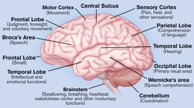
The cerebrum is the largest part of the human brain and is essential for awareness, language, cognition, memory, and consciousness. It is divided into four lobes: frontal, temporal, occipital, and parietal.
Frontal Lobe: Decision making, voluntary movement, and executive functions.
Parietal Lobe: Sensory processing.
Occipital Lobe: Visual processing.
Temporal Lobe: Hearing and language.
Lateralization of Cortical Function
The left and right hemispheres of the brain have specialized functions, a phenomenon known as lateralization. The corpus callosum connects the hemispheres, allowing communication between them.
Information Processing in the Brain
The thalamus directs sensory information to appropriate areas of the cortex. The somatosensory and motor cortices are organized according to body regions.
Concept 4: Changes in Synaptic Connections Underlie Memory and Learning
Neural Plasticity and Memory
Neural plasticity is the ability of the nervous system to change in response to experience. Memory formation involves changes in synaptic strength.
Short-term Memory: Temporary links in the hippocampus.
Long-term Memory: Stored in the cerebral cortex; consolidation occurs during sleep.
Long-Term Potentiation (LTP)
LTP is a long-lasting increase in synaptic strength, involving simultaneous activity in pre- and post-synaptic neurons and the neurotransmitter glutamate.
Concept 5: Nervous System Disorders Can Be Explained in Molecular Terms
Major Nervous System Disorders
Schizophrenia: Affects thought processes and emotional responsiveness.
Depression: Characterized by persistent sadness and loss of interest.
Drug Addiction: Involves changes in the brain's reward system.
Alzheimer’s Disease: Marked by confusion and memory loss, caused by amyloid plaques and neurofibrillary tangles.
Parkinson’s Disease: A motor disorder due to the death of dopamine-producing neurons in the midbrain, leading to tremors and poor balance.
Genetic and environmental factors contribute to these diseases, and treatments often focus on symptom management rather than cures.
Example: Alzheimer’s disease is associated with decreased acetylcholine (ACh) levels, while Parkinson’s disease involves dopamine (DA) and ACh imbalances.
