 Back
BackNervous Systems: Structure, Function, and Disorders
Study Guide - Smart Notes
 Tailored notes based on your materials, expanded with key definitions, examples, and context.
Lecture 15
Nervous System Overview
Lines of Communication
The nervous system is composed of specialized cells called neurons that transfer information throughout the body. Neurons communicate using both electrical signals (for long-distance transmission) and chemical signals (for short-distance communication). Information processing occurs in clusters of neurons called ganglia or in more complex structures such as the brain.
Neurons: Nerve cells responsible for information transfer.
Electrical signals: Used for rapid, long-distance communication within neurons.
Chemical signals: Used for communication between neurons at synapses.
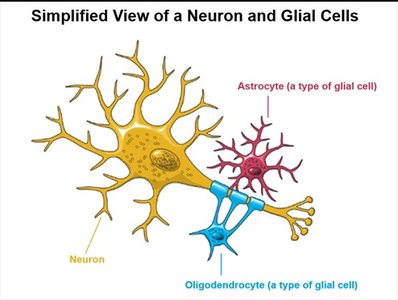
Glial cells: Support and protect neurons in both the CNS and PNS.
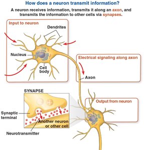
Neuron Structure and Function
Basic Anatomy of a Neuron
Neurons have a unique structure that supports their function in information transfer:
Cell body (soma): Contains the nucleus and most organelles.
Dendrites: Highly branched extensions that receive signals from other neurons.
Axon: A long extension that transmits signals to other cells at synapses.
Axon hillock: The cone-shaped base of the axon where action potentials are initiated.
Synapse: The junction between an axon and another cell, where neurotransmitters are released.
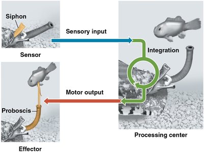
Information Processing in Nervous Systems
Stages of Information Processing
Nervous systems process information in three main stages:
Sensory input: Sensors detect external or internal stimuli and transmit information via sensory neurons.
Integration: Interneurons in the brain or ganglia analyze and interpret sensory information.
Motor output: Motor neurons transmit signals to muscles or glands, triggering a response.
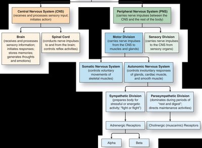
Organization of Nervous Systems
Central and Peripheral Nervous Systems
The nervous system is divided into the central nervous system (CNS) and the peripheral nervous system (PNS):
CNS: Includes the brain and spinal cord; responsible for integration and processing of information.
PNS: Transmits information to and from the CNS; includes sensory and motor divisions.
Somatic nervous system: Controls voluntary movements of skeletal muscles.
Autonomic nervous system: Regulates involuntary functions (e.g., heart rate, digestion).
Sympathetic division: Prepares the body for "fight or flight" responses.
Parasympathetic division: Promotes "rest and digest" activities.
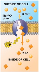
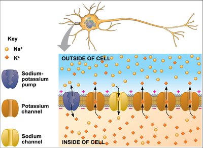
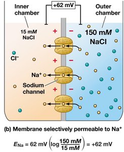
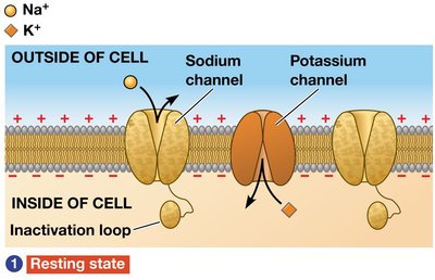
Resting Potential and Ion Channels
Establishing Resting Potential
The resting potential is the membrane potential of a neuron not sending signals. It is established by the action of ion pumps and ion channels:
Sodium-potassium pump: Uses ATP to move 3 Na+ ions out and 2 K+ ions into the cell, maintaining concentration gradients.
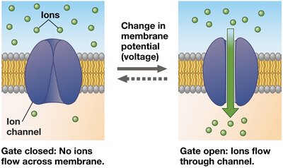
Ion channels: Allow selective movement of ions across the membrane, converting chemical potential to electrical potential.
Modeling Resting Potential
The Nernst equation calculates the equilibrium potential for a particular ion:
At equilibrium, both electrical and chemical gradients are balanced, and the resting potential remains steady.
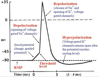
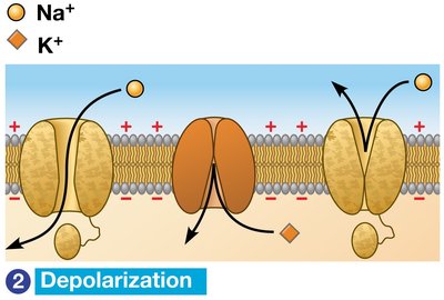
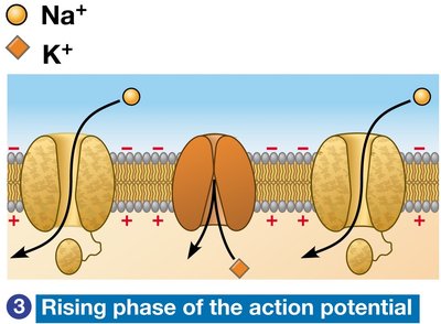
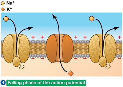
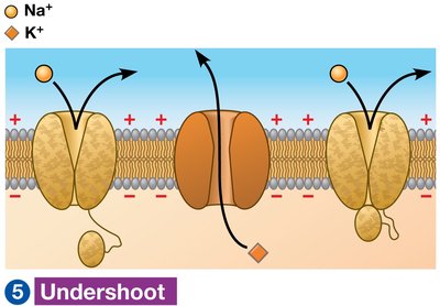
Action Potentials
Generation and Propagation
Action potentials are rapid changes in membrane potential that travel along axons. They are generated by the opening and closing of voltage-gated ion channels in response to stimuli.
Depolarization: Voltage-gated Na+ channels open, Na+ enters the cell.
Rising phase: Membrane potential approaches ENa.
Falling phase: Na+ channels inactivate, K+ channels open, K+ exits the cell.
Undershoot: K+ permeability is high, then returns to resting state.
Refractory period: Na+ channels are temporarily inactivated, preventing backward propagation.
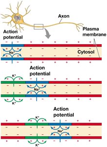
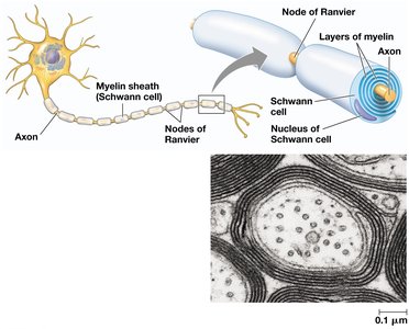
Conduction of Action Potentials
Propagation Along the Axon
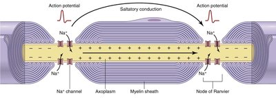
Action potentials are conducted along the axon by the movement of ions. The rate of action potential propagation depends on axon diameter and myelination.
Unmyelinated axons: Conduct impulses slowly and continuously.
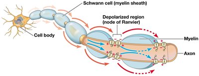
Myelinated axons: Conduct impulses rapidly via saltatory conduction, where the action potential jumps from node to node (nodes of Ranvier).
Myelin sheath: Insulating layer produced by glial cells (oligodendrocytes in CNS, Schwann cells in PNS).
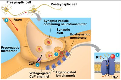
Synaptic Transmission
Structure and Function of Synapses
At synapses, the arrival of an action potential triggers the release of neurotransmitters from the presynaptic neuron. These chemicals cross the synaptic cleft and bind to receptors on the postsynaptic cell, generating a response.
Voltage-gated Ca2+ channels: Open in response to action potential, allowing Ca2+ influx.
Synaptic vesicles: Fuse with the presynaptic membrane to release neurotransmitters.
Ligand-gated ion channels: On the postsynaptic membrane, mediate the postsynaptic potential.
Neurotransmitters and Receptors
Types of Neurotransmitters
Acetylcholine (ACh): Involved in muscle stimulation, memory, and learning; acts on both nicotinic (ionotropic) and muscarinic (metabotropic) receptors.
Amino acids: Glutamate (excitatory), glycine, and GABA (inhibitory).
Biogenic amines: Dopamine, serotonin, norepinephrine, and epinephrine; involved in mood, reward, and autonomic functions.
Neuropeptides: Substance P, endorphins; modulate pain and other functions.
Gases: Nitric oxide (NO), carbon monoxide (CO); act as local regulators.
Neurotransmitter Receptors
Ionotropic receptors: Ligand-gated ion channels that mediate rapid responses (e.g., nicotinic ACh receptors).
Metabotropic receptors: G-protein-coupled receptors that trigger slower, longer-lasting effects (e.g., muscarinic ACh, serotonin, dopamine receptors).
Organization of the Vertebrate Nervous System
Central and Peripheral Components
The vertebrate nervous system is organized into the CNS (brain and spinal cord) and PNS (nerves and ganglia). The CNS develops from the dorsal hollow nerve cord, and its central canal and ventricles are filled with cerebrospinal fluid (CSF).
Gray matter: Neuron cell bodies, dendrites, and unmyelinated axons.
White matter: Bundles of myelinated axons.
Major Brain Regions
Forebrain: Olfactory processing, sleep, learning, complex processing.
Midbrain: Sensory input routing.
Hindbrain: Involuntary activities, motor coordination.
Brain Function and Disorders
Cerebral Cortex and Lateralization
The cerebral cortex is responsible for voluntary movement, cognition, memory, and awareness. The two hemispheres of the brain have specialized functions but communicate via the corpus callosum.
Left hemisphere: Language, math, logic.
Right hemisphere: Pattern recognition, spatial relations, nonverbal thinking.
Neuronal Plasticity
Neuronal plasticity refers to the brain's ability to reorganize neural pathways based on experience, which underlies learning and memory.
Nervous System Disorders
Schizophrenia: Chronic psychiatric disorder involving dopamine dysregulation, genetic and environmental factors.
Alzheimer’s Disease: Dementia characterized by neurofibrillary tangles (tau protein) and amyloid plaques; no cure.
Parkinson’s Disease: Motor disorder due to loss of dopamine-secreting neurons; treated with L-dopa.
Addiction: Drugs increase dopamine activity in the brain's reward system, leading to compulsive use and long-term changes in neural circuitry.
Additional info: This guide covers the structure and function of neurons, mechanisms of action potentials and synaptic transmission, organization of the nervous system, and major neurological disorders, providing a comprehensive overview suitable for college-level biology students.
