 Back
BackNervous Systems: Structure, Function, and Specialization
Study Guide - Smart Notes
 Tailored notes based on your materials, expanded with key definitions, examples, and context.
Nervous System Organization
Central and Peripheral Nervous Systems
The vertebrate nervous system is divided into the central nervous system (CNS) and peripheral nervous system (PNS). Each system has specialized functions and distinct anatomical features.
CNS: Composed of the brain and spinal cord; responsible for processing and integrating information.
PNS: Composed of nerves and ganglia outside the CNS; transmits information to and from the CNS.
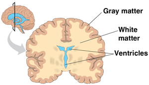
Gray matter: Contains neuron cell bodies, dendrites, and unmyelinated axons.
White matter: Consists of bundles of myelinated axons, enabling rapid communication between regions.
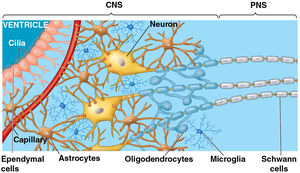
Glial Cells and Neurons
Glial cells (or glia) nourish, support, and regulate neurons. They play essential roles in brain development, maintenance, and repair.
Radial glia: Guide neuron migration during brain development.
Astrocytes: Help form the blood–brain barrier, controlling entry into the brain.
Oligodendrocytes (CNS) and Schwann cells (PNS): Provide myelin sheaths for axons, increasing signal speed.
Microglia: Immune cells of the CNS, removing debris and pathogens.
Unlike most neurons, many glia can divide and self-renew, making them more susceptible to oncogenic mutations (brain tumors).
Functional Anatomy of the Nervous System
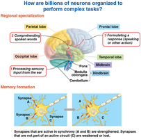
Neuroanatomy and Regional Specialization
Neuroanatomy links brain regions to specific functions, aiding clinical diagnosis and understanding of behavior.
The spinal cord processes reflexes and basic movement, carrying information between the brain and body.
Different brain regions process distinct functions, such as vision, hearing, and language.
Major Brain Regions
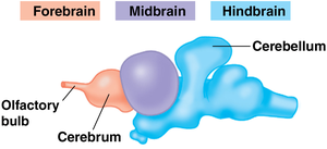
The brain is divided into three main regions, each with specialized functions:
Forebrain: Complex processing, learning, sleep regulation, and olfaction.
Midbrain: Routes sensory information and supports sensorimotor integration.
Hindbrain: Controls involuntary functions and coordinates movement.
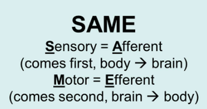
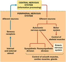
Peripheral Nervous System: Afferent and Efferent Pathways
Afferent vs Efferent Neurons
The PNS transmits information to and from the CNS, regulating movement and the internal environment.
Afferent neurons: Carry sensory information from the body to the CNS.
Efferent neurons: Carry motor commands from the CNS to the body.
Motor Output and Autonomic Nervous System
Efferent pathways are divided into the motor system and autonomic nervous system:
Motor system: Controls skeletal muscle (voluntary movement).
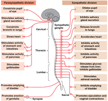
Autonomic nervous system: Controls smooth and cardiac muscle (involuntary functions).
Sympathetic division: Mobilizes energy for "fight-or-flight" responses.
Parasympathetic division: Promotes rest and digestion.
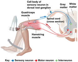
Neural Circuits and Reflexes
Reflex Arcs
The spinal cord is a processing center for reflexes, enabling rapid responses to stimuli without conscious input.
Reflex arcs involve sensory neurons, interneurons, and motor neurons.
Example: Knee-jerk reflex, where sensory input triggers motor output via the spinal cord.
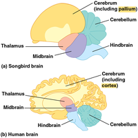
Brain Structure and Evolution
Brain Structure Reflects Function
The size and structure of brain regions reflect their importance and function within the nervous system.
Larger regions perform more critical or heavily used functions.
Example: Sharks have enlarged olfactory regions for smell-guided behavior.
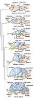
Evolution of Cognition in Vertebrates
The pallium is the outer brain region for complex processing, present in the common ancestor of birds and mammals.
Birds: Clustered pallium for sophisticated cognition.
Mammals: Layered pallium (packs more tissue into less space).
Connectivity preserved, enabling complex cognition in both groups.
Memory Formation and Synaptic Plasticity
Memory Formation
Memory is formed through changes in neural connections, a process known as neuronal plasticity.
Synapses that are active strengthen their connections.
Inactive synapses weaken or are lost.
Memory consolidation involves the hippocampus and cerebral cortex.
Long-Term Potentiation (LTP)
LTP is a lasting increase in synaptic strength, serving as a cellular mechanism for memory formation.
Involves glutamate receptors on the postsynaptic neuron.
Receptor changes make the neuron more responsive to future signals.
Result: The connection becomes stronger and easier to activate.
Equation:
Where is the change in synaptic weight, is the learning rate, is presynaptic activity, and is postsynaptic activity.
Summary Table: Nervous System Divisions and Functions
Division | Main Function | Key Components |
|---|---|---|
CNS | Integration and processing | Brain, spinal cord |
PNS | Transmission of information | Nerves, ganglia |
Motor System | Voluntary movement | Skeletal muscle |
Autonomic System | Involuntary functions | Smooth/cardiac muscle, glands |
Sympathetic Division | Mobilizes energy | Fight-or-flight |
Parasympathetic Division | Promotes rest | Rest and digest |
Additional info:
Neuroanatomy is essential for understanding clinical conditions such as brain injuries, tumors, and neurodegenerative diseases.
Synaptic plasticity underlies learning, memory, and adaptation to new experiences.
