 Back
BackNeurobiology: Synaptic Transmission and Neurotransmitters
Study Guide - Smart Notes
 Tailored notes based on your materials, expanded with key definitions, examples, and context.
Neurobiology: Synaptic Transmission and Neurotransmitters
Neurons and Synaptic Communication
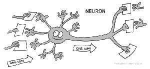
Neurons are specialized cells in the nervous system that transmit information through electrical and chemical signals. Communication between neurons occurs at junctions called synapses, which are critical for neural function and information processing.
Synapse: The junction between two neurons where information is transmitted.
Presynaptic neuron: The neuron sending the signal.
Postsynaptic neuron: The neuron receiving the signal.
Types of synapses: Electrical and chemical synapses.
Electrical synapse: Direct flow of electrical current via gap junctions.
Chemical synapse: Transmission via neurotransmitter release.
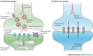
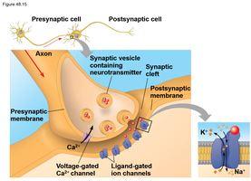
Mechanism of Chemical Synaptic Transmission
Most synapses in the nervous system are chemical synapses. Here, the presynaptic neuron releases neurotransmitters into the synaptic cleft, which bind to receptors on the postsynaptic membrane, triggering a response.
Neurotransmitter: Chemical messenger released from synaptic vesicles.
Synaptic cleft: The gap between presynaptic and postsynaptic neurons.
Receptors: Proteins on the postsynaptic membrane that bind neurotransmitters.
Ion channels: Open or close in response to neurotransmitter binding, altering membrane potential.
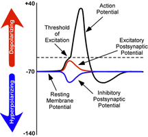
Postsynaptic Potentials: EPSP and IPSP
Binding of neurotransmitters to postsynaptic receptors generates postsynaptic potentials, which are changes in membrane potential. These can be excitatory or inhibitory.
Excitatory Postsynaptic Potential (EPSP): Depolarizes the membrane, increasing the likelihood of action potential.
Inhibitory Postsynaptic Potential (IPSP): Hyperpolarizes the membrane, decreasing the likelihood of action potential.
Threshold: The membrane potential at which an action potential is triggered.
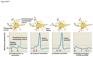
Summation of Postsynaptic Potentials
Neurons integrate multiple EPSPs and IPSPs through summation, which determines whether an action potential will occur. Summation can be temporal (over time) or spatial (over multiple synapses).
Temporal summation: Multiple signals from one synapse in rapid succession.
Spatial summation: Signals from multiple synapses at the same time.
Integration: The process by which the neuron decides to fire an action potential based on combined inputs.


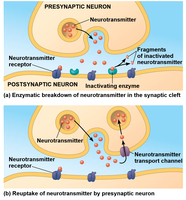
Termination of Neurotransmitter Signaling
Neurotransmitter signaling must be terminated to prevent continuous stimulation. This occurs through enzymatic breakdown or reuptake by the presynaptic neuron.
Enzymatic breakdown: Enzymes in the synaptic cleft degrade neurotransmitters.
Reuptake: Neurotransmitters are transported back into the presynaptic neuron.
Example: Acetylcholine is broken down by acetylcholinesterase.
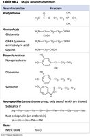
Major Neurotransmitters
Neurotransmitters are classified by their chemical structure and function. Major classes include amino acids, biogenic amines, neuropeptides, and gases.
Amino acids: Glutamate, GABA, glycine.
Biogenic amines: Dopamine, serotonin, norepinephrine.
Neuropeptides: Substance P, endorphins.
Gases: Nitric oxide.
Neurotransmitter | Structure |
|---|---|
Acetylcholine | CH3COOCH2CH2N+(CH3)3 |
Glutamate | HOOC(CH2)2CH(NH2)COOH |
GABA | NH2CH2CH2CH2COOH |
Glycine | NH2CH2COOH |
Norepinephrine | Additional info: Catecholamine structure |
Dopamine | Additional info: Catecholamine structure |
Serotonin | Additional info: Indoleamine structure |
Substance P | Peptide |
Endorphin | Peptide |
Nitric oxide | NO |
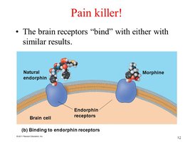
Neuropeptides and Pain Modulation
Neuropeptides are small protein-like molecules used by neurons to communicate. Endorphins are neuropeptides that act as natural painkillers by binding to opioid receptors in the brain.
Endorphins: Inhibit pain by binding to receptors also targeted by morphine.
Example: Morphine mimics endorphin action, providing pain relief.
Biogenic Amines and Psychotropic Medication
Biogenic amines such as serotonin, norepinephrine, and dopamine are involved in mood regulation and are targets for psychotropic medications. Drugs like antidepressants and Ritalin modulate neurotransmitter-mediated transmission at synapses.
Antidepressants: Often inhibit reuptake of serotonin or norepinephrine, increasing their levels in the synaptic cleft.
Ritalin: Modulates dopamine transmission, used in ADHD treatment.
Mechanism: Drugs alter neurotransmitter release, reuptake, or degradation.


Summary Table: Neurotransmitter Classes
The following table summarizes the main classes of neurotransmitters, their examples, and primary functions.
Class | Example | Function |
|---|---|---|
Amino Acid | Glutamate, GABA | Excitatory/inhibitory signaling |
Biogenic Amine | Dopamine, Serotonin | Mood, reward, arousal |
Neuropeptide | Endorphin, Substance P | Pain modulation, signaling |
Gas | Nitric oxide | Modulation of blood flow |
Additional info:
Neurobiology is covered in Chapter 48 of most biology textbooks, focusing on neurons, synapses, and neurotransmitter function.
Psychotropic drugs are discussed in the context of neurotransmitter modulation and their effects on mental health.
