 Back
Lecture 6 Neurons, Synapses, and Kidney Regulation: Structure, Function, and Comparative Physiology
Study Guide - Smart Notes
 Tailored notes based on your materials, expanded with key definitions, examples, and context.
Lecture 6 Kidney Regulation & Comparative Physiology
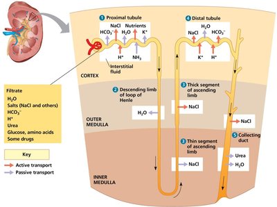
Overview of Nephron Structure and Function
The nephron is the functional unit of the kidney, responsible for filtering blood and forming urine. Its structure and function are adapted to the osmoregulatory needs of different vertebrates.
Filtration: Occurs in the glomerulus, producing a filtrate containing water, ions, glucose, amino acids, and waste products.
Reabsorption: Essential solutes and water are reabsorbed along the nephron, especially in the proximal tubule and loop of Henle.
Secretion: Additional wastes and ions are secreted into the filtrate in the distal tubule and collecting duct.
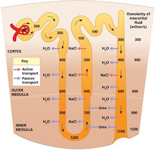
Osmotic Gradient: Urea and NaCl establish an osmotic gradient in the medulla, enabling the production of urine that is hyperosmotic to blood.
Osmolarity and Countercurrent Multiplication
The osmolarity of the interstitial fluid increases from the cortex to the inner medulla, facilitating water reabsorption and urine concentration.
Descending Limb of Loop of Henle: Permeable to water but not to NaCl; water exits, concentrating the filtrate.
Ascending Limb of Loop of Henle: Impermeable to water; NaCl is actively and passively transported out, diluting the filtrate.
Collecting Duct: Regulated reabsorption of water and urea, final adjustment of urine concentration.
Hormonal Regulation of Kidney Function
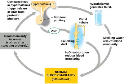
Three major hormonal systems regulate water and salt balance in the kidney: Antidiuretic Hormone (ADH), the Renin–Angiotensin–Aldosterone System (RAAS), and Atrial Natriuretic Peptide (ANP).
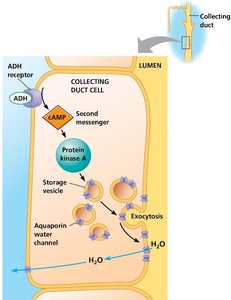
ADH (Vasopressin): Released in response to increased blood osmolarity; increases water reabsorption in the collecting duct by promoting aquaporin insertion.
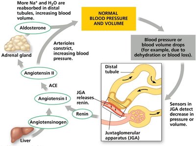
RAAS: Activated by decreased blood pressure/volume; increases Na+ and water reabsorption via aldosterone, raising blood pressure and volume.
ANP: Released in response to increased blood volume; opposes RAAS and ADH, promoting natriuresis and diuresis.
ADH Mechanism of Action
ADH binds to receptors on collecting duct cells, triggering a cAMP-mediated pathway that results in the insertion of aquaporin water channels into the apical membrane, increasing water permeability and reabsorption.
Effect: Concentrates urine and reduces blood osmolarity.
Clinical Note: Mutations affecting ADH or aquaporin can cause diabetes insipidus, characterized by excessive urination and dehydration.
RAAS and Aldosterone Mechanism
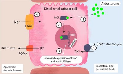
The RAAS system is activated by low blood pressure or volume. Renin release leads to the formation of angiotensin II, which constricts arterioles and stimulates aldosterone secretion from the adrenal cortex. Aldosterone increases Na+ reabsorption and K+ secretion in the distal tubule and collecting duct, raising blood volume and pressure.
Time Scale: ADH acts within minutes; aldosterone acts over hours to days.
Mechanism: ADH acts via membrane GPCRs; aldosterone acts via intracellular receptors to alter gene expression (e.g., ENaC, Na+/K+-ATPase).
Comparative Kidney Structure and Adaptations
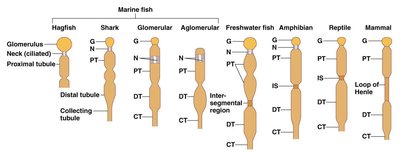
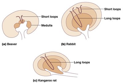
Kidney structure varies among vertebrates, reflecting adaptations to different environments. Mammals in arid habitats have longer loops of Henle for greater water conservation, while aquatic species have shorter loops.
Reniculated Kidneys: Found in marine mammals and large terrestrial mammals, composed of multiple lobes for increased filtration and processing capacity.
Hibernation Adaptations: Bears recycle urea during hibernation, minimizing water loss and kidney damage.
Animal Electricity: Historical Perspective
Discovery of Animal Electricity
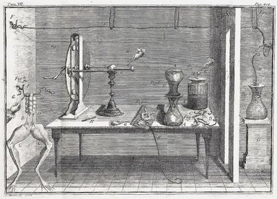
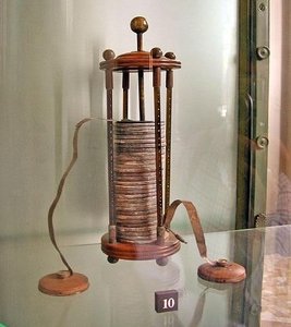
The concept of animal electricity originated from experiments in the late 18th century, where electrical phenomena were observed in dissected frog muscles. Luigi Galvani proposed that nerves and muscles generate intrinsic electrical signals, while Alessandro Volta argued that electricity was produced by contact between different metals.
Galvani's Experiments: Demonstrated muscle contraction in frog legs upon electrical stimulation.
Volta's Battery: Showed that dissimilar metals and electrolytes could generate electricity, leading to the invention of the voltaic pile (battery).
Neurons, Synapses, and Signaling
Membrane Potential and Action Potentials
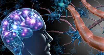
Neurons transmit information via changes in membrane potential. The resting membrane potential is maintained by ion gradients, primarily through the action of the Na+/K+-ATPase pump. Action potentials are rapid, all-or-none changes in membrane voltage that propagate along axons.
Resting Potential: Typically -65 to -80 mV, maintained by selective permeability and active transport of ions.
Action Potential: Triggered by depolarization above threshold, involving voltage-gated Na+ and K+ channels.
Refractory Period: Ensures unidirectional propagation of action potentials.
Myelination: Increases conduction speed via saltatory conduction at nodes of Ranvier.
Neuronal Communication: Synapses
Neurons communicate at synapses, which can be electrical (via gap junctions) or chemical (via neurotransmitter release). Chemical synapses involve the release of neurotransmitters from presynaptic vesicles, which bind to receptors on the postsynaptic cell, generating excitatory or inhibitory postsynaptic potentials (EPSPs or IPSPs).
Ionotropic Receptors: Ligand-gated ion channels that mediate fast synaptic transmission.
Metabotropic Receptors: G-protein coupled receptors that activate second messenger pathways, producing slower but longer-lasting effects.
Summation: Temporal and spatial summation of EPSPs and IPSPs determines whether the postsynaptic neuron reaches threshold for action potential generation.
Key Equations and Concepts
Ohm's Law:
Time Constant (\tau):
Equilibrium Potential (Nernst Equation):
Examples and Applications
Diabetes Insipidus: Caused by mutations in ADH or aquaporin genes, leading to excessive urination and dehydration.
Puffer Fish Toxin: Tetrodotoxin blocks voltage-gated Na+ channels, preventing action potential generation and causing paralysis.
Additional info: This guide integrates textbook diagrams and experimental data to reinforce understanding of kidney physiology, neuronal signaling, and the historical development of electrophysiology.
