 Back
L_8 Neurons, Synapses, and Sensory Systems: Structure and Function of the Nervous System
Study Guide - Smart Notes
 Tailored notes based on your materials, expanded with key definitions, examples, and context.
L_8 Neurons, Synapses, and Signaling
Saltatory Conduction and Synaptic Transmission
The nervous system relies on specialized cells called neurons to transmit information rapidly throughout the body. Action potentials (APs) propagate along axons, and in myelinated neurons, this process is called saltatory conduction, where the AP 'jumps' between nodes of Ranvier, increasing conduction speed. Communication between neurons occurs at synapses, where neurotransmitters are released to transmit signals to the next cell.
Comparative Organization of Nervous Systems
Evolution and Development of the Human Brain
The vertebrate nervous system is organized into central and peripheral components. During embryonic development, the neural tube forms three primary brain vesicles: forebrain, midbrain, and hindbrain. These regions further differentiate into specialized structures in the adult brain, each with distinct functions.
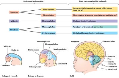
Structure and Function of the Human Brain
The Cerebrum and Cerebral Cortex
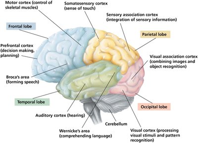
The cerebrum is the largest part of the human brain and is essential for higher-order functions such as awareness, language, cognition, memory, and consciousness. The cerebral cortex is divided into four lobes, each associated with specific functions:
Frontal lobe: Decision making, planning, voluntary movement, and executive functions.
Parietal lobe: Sensory integration, touch, and spatial orientation.
Temporal lobe: Hearing, language comprehension, and memory.
Occipital lobe: Visual processing.
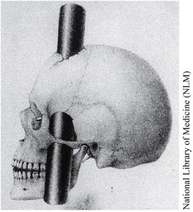
Frontal Lobe Function and Phineas Gage
Damage to the frontal lobe can impair decision making and emotional responses while leaving intellect and memory intact. The famous case of Phineas Gage, who survived a traumatic injury to his frontal lobe, demonstrated the role of this region in personality and executive function.
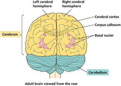
Hemispheres, Basal Nuclei, and Corpus Callosum
The cerebrum consists of right and left hemispheres connected by the corpus callosum. Deep within the white matter are the basal nuclei, which are involved in motor planning and coordination.
Gray Matter, White Matter, and Cerebrospinal Fluid (CSF)
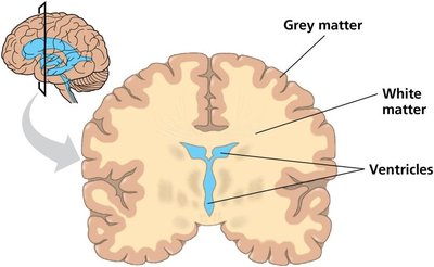
The brain and spinal cord contain gray matter (neuron cell bodies, dendrites, unmyelinated axons) and white matter (myelinated axons). The central canal and ventricles are filled with cerebrospinal fluid, which cushions the brain, provides nutrients, and removes wastes.
Major Brain Divisions and Functions
Diencephalon
Hypothalamus: Regulates homeostasis, hormone release, appetite, and circadian rhythms.
Pineal gland: Releases melatonin, regulating diurnal rhythms.
Thalamus: Relay center for sensory information.
Mesencephalon (Midbrain)
Ventral Tegmental Area (VTA): Reward and motivation (dopamine pathways).
Periaqueductal Gray (PAG): Pain modulation.
Substantia Nigra: Dopamine production; degeneration leads to Parkinson’s Disease.
Metencephalon and Myelencephalon
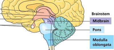
Pons: Relays signals, regulates sleep, respiration, and arousal (serotonin and norepinephrine sources).
Cerebellum: Motor coordination and learning.
Medulla oblongata: Controls vital autonomic functions (breathing, heart rate, blood pressure) and protective reflexes.
Sensory Systems and Sensory Coding
Overview of Sensory Systems
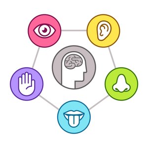
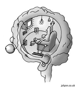
Sensory systems detect and encode environmental stimuli, allowing organisms to perceive and respond to their surroundings. The five main senses are vision, hearing, touch, taste, and smell.
Encoding Sensations in Spike Trains
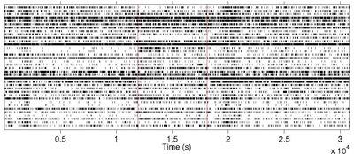
Neurons encode sensory information as patterns of action potentials (spike trains). Different neurons and firing patterns represent different sensory modalities and stimulus intensities.
Evolution and Diversity of Vision
Light Detection in Animals
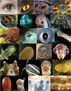
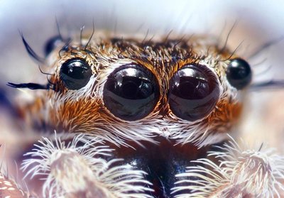
Animals use a variety of organs for vision, but all rely on photoreceptors containing light-absorbing pigments. Vision systems range from simple eyespots (ocelli) to complex image-forming eyes.
Ocelli and Orientation in Planarians
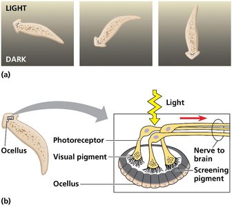
Planarians possess simple eyespots (ocelli) that detect light direction and intensity, allowing them to move away from light and seek shaded environments.
Compound Eyes
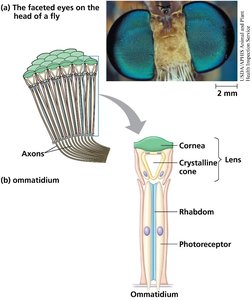
Insects and crustaceans have compound eyes composed of many ommatidia, each functioning as a separate light detector. Compound eyes are highly effective at detecting movement and, in some species, color and ultraviolet light.
Single-Lens Eyes
Single-lens eyes, found in vertebrates and some invertebrates, function like a camera. The iris controls the amount of light entering the eye, and the lens focuses light onto the retina.
The Vertebrate Visual System
Vertebrate eyes detect color and light for both visual and non-visual functions (e.g., circadian rhythms). Photoreceptors in the retina convert light into electrical signals, which are processed and relayed to the brain.
Retinal Structure and Visual Processing
Distribution of Photoreceptors
The human retina contains approximately 5 million cones (for color vision) concentrated in the fovea, and a larger number of rods (for low-light vision). Saccadic eye movements allow the fovea to sample different parts of the visual field.
Color Vision and Retinal Circuitry
Humans perceive color using three types of cones (red, green, blue), each with a distinct opsin protein. Retinal circuitry involves five types of neurons: photoreceptors, bipolar cells, ganglion cells, horizontal cells, and amacrine cells. Lateral inhibition enhances contrast and edge detection.
Visual Pathways
Visual information from the left visual field is processed by the right hemisphere and vice versa. Most ganglion cell axons project to the lateral geniculate nucleus, which relays information to the primary visual cortex.
Types of Sensory Receptors
Classification of Sensory Receptors
Sensory receptors are classified by the type of stimulus energy they transduce:
Mechanoreceptors: Detect touch, pressure, vibration, and stretch.
Chemoreceptors: Detect chemical stimuli (taste, smell, blood pH).
Electromagnetic receptors: Detect light and electromagnetic fields.
Thermoreceptors: Detect temperature changes.
Mechanoreceptors and Somatosensory Pathways
Mechanoreceptors sense physical deformation. The cell bodies of primary sensory neurons are located in the dorsal root ganglia (DRG). Sensory information is relayed through the spinal cord and thalamus to the somatosensory cortex.
Touch Receptors and Sensory Maps
Touch receptors are unevenly distributed across the body, resulting in varying sensitivity. The two-point discrimination threshold measures receptor density. The somatosensory cortex contains a 'homunculus' map reflecting receptor distribution.
Nociception and Pain Perception
Mechanoreception vs. Nociception
Nociceptors detect harmful stimuli (excess heat, pressure, chemicals) and are distinct from other sensory receptors. Nociception refers to the sensory detection of noxious stimuli, while pain is the subjective, conscious experience.
Hearing and the Auditory System
Structure and Function of the Ear
The outer ear amplifies sound waves, which cause vibration of the eardrum. The Organ of Corti in the cochlea contains hair cells that transduce mechanical energy into electrical signals, which are sent to the brain via the auditory nerve.
Chemical Senses: Taste and Smell
Taste (Gustation)
Taste receptor cells are organized into taste buds and detect five basic tastes: sweet, sour, salty, bitter, and umami. Different receptor types (GPCRs, TRP channels, sodium channels) mediate these sensations.
Smell (Olfaction)
Olfactory receptor neurons in the nasal cavity each express a single type of GPCR. Odor perception is based on a combinatorial code, allowing mammals to distinguish thousands of odors.
Summary Table: Major Brain Regions and Functions
Brain Region | Main Structures | Primary Functions |
|---|---|---|
Forebrain | Cerebrum, Diencephalon | Higher cognition, sensory processing, homeostasis, hormone regulation |
Midbrain | Mesencephalon | Motor control, reward, sensory integration |
Hindbrain | Pons, Cerebellum, Medulla oblongata | Motor coordination, autonomic control, signal relay |
