 Back
BackNeurons, Synapses, and Signaling: Foundations of Nervous System Function
Study Guide - Smart Notes
 Tailored notes based on your materials, expanded with key definitions, examples, and context.
Neurons, Synapses, and Signaling
Introduction to Neurons and Nervous System Function
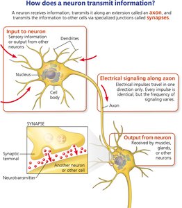
The nervous system is composed of specialized cells called neurons that transmit information using electrical and chemical signals. Neurons are the fundamental units of the nervous system, responsible for receiving, integrating, and transmitting information throughout the body. Groups of neurons form complex circuits that underlie all nervous system functions, including sensation, movement, and cognition.
Neurons use electrical impulses (action potentials) to transmit information over long distances.
Communication between neurons occurs at specialized junctions called synapses, where chemical messengers called neurotransmitters relay signals to other cells.
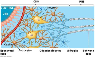
Neural circuits are organized into the central nervous system (CNS) and peripheral nervous system (PNS).
Neuron Structure and Function
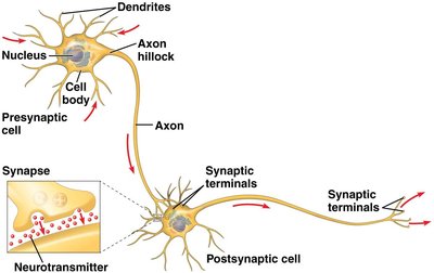
Neurons have a characteristic structure that supports their function in information processing and transmission:
Dendrites: Branched extensions that receive signals from other neurons.
Cell body (soma): Contains the nucleus and integrates incoming signals.
Axon hillock: The region where action potentials are initiated.
Axon: A long projection that conducts action potentials away from the cell body.
Synaptic terminals: The ends of the axon where neurotransmitters are released to communicate with other cells.
Types of Neurons
Neurons are classified based on their function:
Sensory neurons: Transmit information about external and internal stimuli to the CNS.
Interneurons: Integrate information within the CNS; most neurons in the brain are interneurons.
Motor neurons: Transmit signals from the CNS to muscles or glands, triggering a response.
Supporting Cells: Glia
Glial cells (or glia) support neuron function by nourishing neurons, insulating axons, maintaining the extracellular environment, and participating in immune defense and nervous system development.
Electrical Properties of Neurons
Resting Membrane Potential
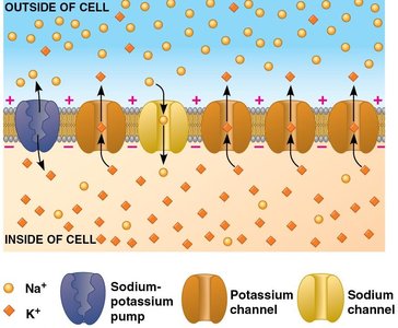
The resting potential is the voltage difference across the plasma membrane of a neuron at rest, typically between -60 and -80 mV. This potential is established by the unequal distribution of ions across the membrane and the selective permeability of ion channels.
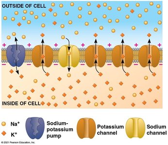
Potassium (K+) is more concentrated inside the cell, while sodium (Na+) is more concentrated outside.
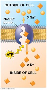
The sodium-potassium pump (Na+/K+-ATPase) maintains these gradients by pumping 3 Na+ out and 2 K+ in, using ATP.
K+ leak channels allow K+ to move out of the cell, making the inside more negative.
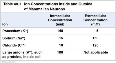
Ion | Intracellular (mM) | Extracellular (mM) |
|---|---|---|
Potassium (K+) | 140 | 5 |
Sodium (Na+) | 15 | 150 |
Chloride (Cl-) | 10 | 120 |
Large anions (A-) | 100 | Not applicable |
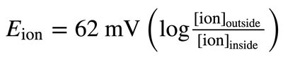
The Nernst Equation
The equilibrium potential for an ion is calculated using the Nernst equation, which predicts the membrane potential at which there is no net movement of a particular ion:
Ion Channels and Membrane Permeability
Ion channels are proteins that allow specific ions to move across the membrane. The resting potential is primarily determined by the membrane's permeability to K+, but a small Na+ leak makes the resting potential less negative than the K+ equilibrium potential.
Action Potentials
Generation and Propagation of Action Potentials
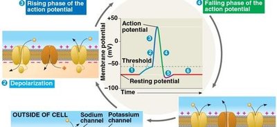
An action potential is a rapid, all-or-none change in membrane potential that travels along the axon. It is triggered when the membrane potential reaches a threshold (about -55 mV in mammals).
Depolarization: Voltage-gated Na+ channels open, Na+ enters the cell, and the membrane potential becomes more positive.
Repolarization: Na+ channels inactivate, K+ channels open, and K+ exits the cell, returning the membrane potential to negative values.
Refractory period: Brief inactivation of Na+ channels prevents the action potential from traveling backward.
Graded Potentials vs. Action Potentials
Graded potentials are small changes in membrane potential that decay with distance and do not trigger an action potential unless they reach threshold. Action potentials are large, self-propagating signals that travel the length of the axon without decreasing in magnitude.
Synaptic Transmission
Chemical Synapses
At chemical synapses, the arrival of an action potential at the synaptic terminal triggers the release of neurotransmitters, which cross the synaptic cleft and bind to receptors on the postsynaptic cell, generating a new signal.
Neurotransmitter release is triggered by Ca2+ influx into the presynaptic terminal.
Neurotransmitters bind to ligand-gated ion channels (ionotropic receptors) or G-protein coupled receptors (metabotropic receptors) on the postsynaptic cell.

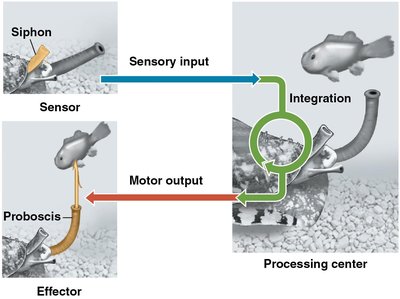
Stages of Information Processing
Nervous system function can be divided into four stages:
Sensory input: Detection of stimuli by sensory receptors.
Integration: Processing and interpretation of sensory input.
Motor output: Transmission of signals to effector cells (muscles or glands).
Learning & Memory: Modification of responses based on experience.
Summary Table: Ion Concentrations in Neurons
Ion | Intracellular Concentration (mM) | Extracellular Concentration (mM) |
|---|---|---|
Potassium (K+) | 140 | 5 |
Sodium (Na+) | 15 | 150 |
Chloride (Cl-) | 10 | 120 |
Large anions (A-) | 100 | Not applicable |
Key Takeaways
Neurons transmit information using electrical impulses and chemical signals.
The resting membrane potential is established by ion gradients and selective permeability.
Action potentials are all-or-none signals that propagate along axons.
Synaptic transmission allows communication between neurons and other cells.
Glial cells support and modulate neuronal function.
