 Back
Neurons, Synapses, and Signaling: Membrane Potentials and Action Potentials
Study Guide - Smart Notes
 Tailored notes based on your materials, expanded with key definitions, examples, and context.
Neurons, Synapses, and Signaling
Overview of Neuronal Function
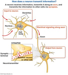
Neurons are specialized cells responsible for receiving, processing, and transmitting information throughout the nervous system. They communicate via electrical impulses and chemical signals, enabling rapid responses to internal and external stimuli.
Neuronal Structure: Neurons consist of dendrites (input), a cell body (integration), and an axon (output).
Synapse: The junction where a neuron communicates with another cell via neurotransmitters.
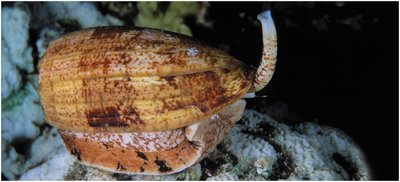
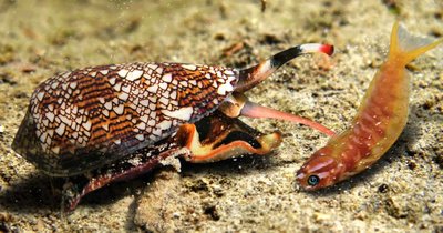
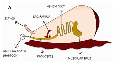
Example: The venomous cone snail (Conus geographus) uses its nervous system to coordinate a rapid, paralyzing attack on prey.
Information Processing in the Nervous System
Three Stages of Information Processing
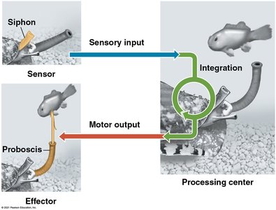
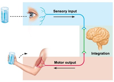
The nervous system processes information in three main stages: sensory input, integration, and motor output. This sequence is fundamental to both invertebrate and vertebrate nervous systems.
Sensory Input: Detection of stimuli by sensory receptors.
Integration: Processing and interpretation of sensory input in the central nervous system.
Motor Output: Activation of effector organs (muscles or glands) in response to integration.
Example: The cone snail senses prey, integrates the information, and triggers a motor response to inject venom.
Membrane Potentials in Neurons
Resting Membrane Potential (RMP)
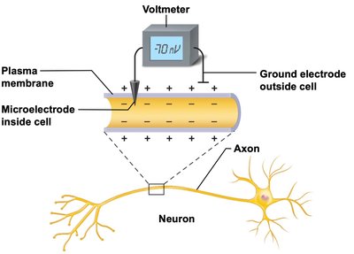
All cells maintain a voltage difference across their plasma membrane, known as the membrane potential. In neurons, the resting membrane potential (RMP) is typically about -70 mV, with the inside of the cell being more negative than the outside.
Polarization: The neuron at rest is polarized due to unequal distribution of ions across the membrane.
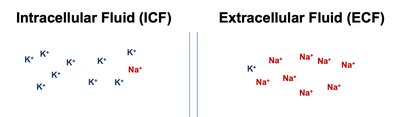
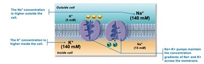
Key Ions: Potassium (K+) is more concentrated inside the cell, while sodium (Na+) is more concentrated outside.
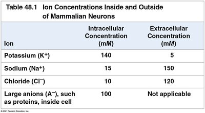
Ion Concentrations Inside and Outside Neurons
The concentration gradients of key ions are essential for establishing the RMP. The following table summarizes the main ion concentrations:
Ion | Intracellular Concentration (mM) | Extracellular Concentration (mM) |
|---|---|---|
Potassium (K+) | 140 | 5 |
Sodium (Na+) | 15 | 150 |
Chloride (Cl-) | 10 | 120 |
Large anions (A-, proteins) | 100 | Not applicable |
Establishing and Maintaining the Resting Membrane Potential
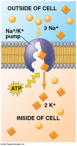
The RMP is established by differences in ion concentration and membrane permeability. The cell membrane is much more permeable to K+ than Na+ due to more K+ leak channels. The Na+/K+ pump actively transports 3 Na+ out and 2 K+ in, maintaining the gradients.
Diffusion Gradients: K+ tends to exit the cell, Na+ tends to enter.
Na+/K+ Pump: Maintains ion gradients using ATP.
Electrical and Chemical Gradients
Ion movement across the membrane is driven by both electrical (voltage) and chemical (concentration) gradients. The equilibrium potential for each ion can be calculated using the Nernst equation:
Nernst Equation:
Typical Values: mV, mV
Additional info: Students are not required to memorize or calculate the Nernst equation for exams, but should understand its conceptual significance.
Membrane Channels and Ion Flow
Types of Membrane Channels
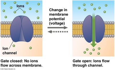
Neurons possess several types of membrane proteins that regulate ion flow:
Leak Channels: Always open, allow passive movement of ions (mainly K+).
Voltage-Gated (VG) Channels: Open or close in response to changes in membrane potential.
Graded Potentials
Definition and Properties
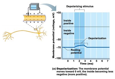
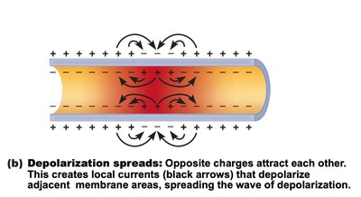
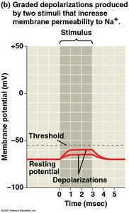
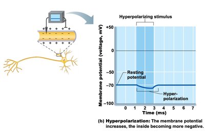
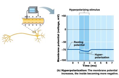
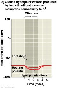
Graded potentials are small, short-lived changes in membrane potential caused by the opening of specific ion channels in response to a stimulus. They can be depolarizing (making the inside less negative) or hyperpolarizing (making the inside more negative).
Depolarization: Usually caused by Na+ influx through VG-Na+ channels.
Hyperpolarization: Usually caused by K+ efflux through VG-K+ channels.
Summation: Multiple graded potentials can add together, increasing the likelihood of reaching threshold for an action potential.
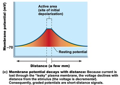
Local Flow: Graded potentials spread locally and decay with distance.
Action Potentials
Graded Potentials vs. Action Potentials
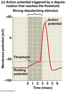
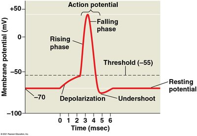
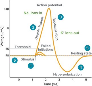
Action potentials are large, rapid, and stereotypical changes in membrane potential that can travel long distances along the axon. They are triggered when a depolarizing graded potential reaches a threshold voltage (typically -50 to -55 mV) at the axon hillock.
All-or-None Principle: Action potentials either occur fully or not at all; their magnitude does not vary with stimulus strength.
Phases of Action Potential:
Stimulus reaches threshold
Depolarization (VG-Na+ channels open, Na+ influx)
Repolarization (VG-K+ channels open, K+ efflux)
Hyperpolarization (K+ channels close slowly)
Return to RMP (Na+/K+ pump restores gradients)
Refractory Periods
After an action potential, the neuron enters a refractory period during which it is less excitable:
Absolute Refractory Period: No new action potential can be initiated (VG channels are inactivated).
Relative Refractory Period: A stronger-than-normal stimulus can initiate another action potential (VG channels are resetting).
Summary Table: Graded Potentials vs. Action Potentials
Feature | Graded Potential | Action Potential |
|---|---|---|
Amplitude | Varies with stimulus strength | All-or-none, fixed size |
Duration | Short, decays with distance | Longer, propagates without decay |
Location | Dendrites, cell body | Axon hillock, axon |
Summation | Possible | Not possible |
Additional info: Understanding the mechanisms of membrane potentials and action potentials is foundational for further study of synaptic transmission, neural circuits, and higher nervous system functions.
