 Back
L7_8 Neurons, Synapses, and Signaling: Structure and Function of the Nervous System
Study Guide - Smart Notes
 Tailored notes based on your materials, expanded with key definitions, examples, and context.
L7_8 Neurons, Synapses, and Signaling
Overview of Nervous System Function
The nervous system is responsible for rapid communication and coordination in animals, allowing them to sense and respond to their environment. Neurons are the fundamental units of the nervous system, transmitting electrical and chemical signals throughout the body.
Neurons: Specialized cells that transmit nerve impulses.
Glia: Support cells that nourish, insulate, and regulate neurons.
Synapses: Junctions where neurons communicate with other cells.
Action Potentials and Nerve Signaling
Resting Membrane Potential
Neurons maintain a resting membrane potential, typically around -70 mV, due to the differential distribution of ions across the plasma membrane. This potential is established by ion channels and the sodium-potassium pump.
Sodium-potassium pump (Na+/K+ ATPase): Actively transports 3 Na+ ions out and 2 K+ ions into the cell, consuming ATP.
Ion channels: Allow passive movement of ions; K+ channels are more permeable at rest.


The equilibrium potentials for K+ and Na+ can be calculated using the Nernst equation:

Generation of Action Potentials
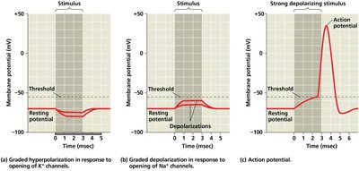
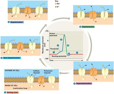
An action potential is a rapid, transient change in membrane potential that travels along the axon. It is triggered when the membrane potential reaches a threshold, usually due to depolarization from incoming signals.
Depolarization: Opening of voltage-gated Na+ channels causes Na+ influx.
Repolarization: Opening of voltage-gated K+ channels causes K+ efflux.
Hyperpolarization: Membrane potential becomes more negative than resting potential before returning to baseline.
Voltage-Gated Ion Channels
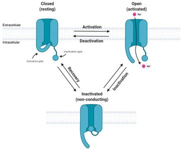
Voltage-gated Na+ and K+ channels are essential for action potential generation and propagation. These channels cycle through closed, open, and inactivated states.
Na+ channels: Open rapidly upon depolarization, then inactivate.
K+ channels: Open more slowly, contributing to repolarization and hyperpolarization.
Propagation of Action Potentials
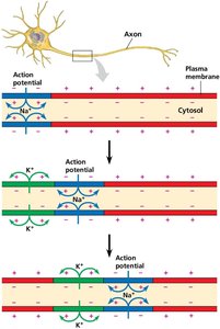
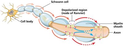
Action potentials propagate along the axon by depolarizing adjacent regions of the membrane. The refractory period ensures unidirectional propagation.
Refractory period: Time during which a neuron cannot fire another action potential, due to inactivated Na+ channels.
Unidirectional conduction: Ensured by refractory period and inactivation of Na+ channels behind the action potential.
Myelination and Saltatory Conduction
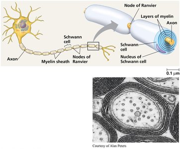
In vertebrates, axons are often wrapped in myelin sheaths, which are produced by Schwann cells (PNS) or oligodendrocytes (CNS). Myelin increases the speed of action potential conduction by insulating the axon and forcing action potentials to "jump" between nodes of Ranvier (saltatory conduction).
Myelin sheath: Insulating layer that increases conduction velocity.
Nodes of Ranvier: Gaps in myelin where voltage-gated channels are concentrated.
Saltatory conduction: Action potentials jump from node to node, increasing speed and efficiency.
Synaptic Transmission
Chemical Synapses
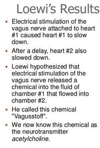
At chemical synapses, an action potential arriving at the presynaptic terminal triggers the release of neurotransmitters, which cross the synaptic cleft and bind to receptors on the postsynaptic cell.
Neurotransmitter release: Triggered by Ca2+ influx into the presynaptic terminal.
Postsynaptic response: Neurotransmitters bind to ligand-gated ion channels, causing excitatory or inhibitory postsynaptic potentials (EPSPs or IPSPs).
Neurotransmitters
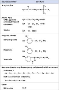
Neurotransmitters are chemical messengers that transmit signals across synapses. They include amino acids, biogenic amines, neuropeptides, and gases.
Examples: Acetylcholine, glutamate, GABA, dopamine, serotonin, substance P, endorphins, nitric oxide.
Clinical relevance: Drugs can affect neurotransmitter levels (e.g., SSRIs, MAO inhibitors).
Neuronal Morphology and Circuitry
Neuronal Structure
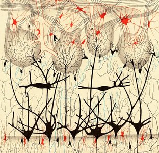
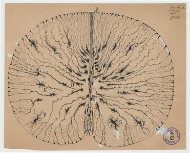
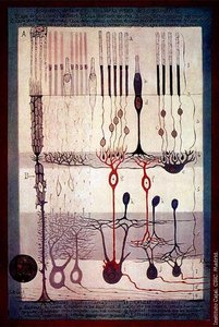
Neurons have diverse morphologies that reflect their functions in neural circuits. Classic studies by Golgi and Ramón y Cajal revealed the intricate architecture of neurons and established the neuron doctrine.
Dendrites: Receive input from other neurons.
Axon: Conducts action potentials away from the cell body.
Synaptic terminals: Release neurotransmitters to communicate with target cells.
Summary Table: Key Ion Channels and Their Roles
Channel Type | Main Ion(s) | Role in Action Potential |
|---|---|---|
Voltage-gated Na+ channel | Na+ | Rapid depolarization (rising phase) |
Voltage-gated K+ channel | K+ | Repolarization and hyperpolarization (falling phase) |
Leak channels | K+ (mainly) | Maintain resting membrane potential |
Na+/K+ ATPase | Na+, K+ | Restores ion gradients after action potentials |
Additional info: The notes above integrate foundational concepts from Campbell Biology, Chapters 48-50, and are suitable for college-level study of the nervous system, focusing on the structure and function of neurons, action potentials, synaptic transmission, and the organization of the nervous system.
