 Back
BackNeurons, Synapses, and Signaling: Structure and Function in the Nervous System
Study Guide - Smart Notes
 Tailored notes based on your materials, expanded with key definitions, examples, and context.
Neurons, Synapses, and Signaling
Neuron Structure and Organization
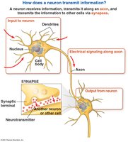
The structure of neurons is specialized for the transfer of information within the nervous system. Neurons are the fundamental units of the nervous system, responsible for receiving, integrating, and transmitting signals.
Dendrites: Branch-like extensions that receive signals from other neurons.
Cell Body (Soma): Contains the nucleus and organelles; integrates incoming signals.
Axon: A long projection that transmits electrical impulses away from the cell body toward other cells.
Axon Hillock: The region where the axon joins the cell body; site of action potential initiation.
Synaptic Terminals: Endings of axons where neurotransmitters are released to communicate with other cells.
Synapse: The junction between a neuron and another cell (neuron, muscle, or gland).
Glia (Glial Cells): Support cells that nourish, insulate, and protect neurons.
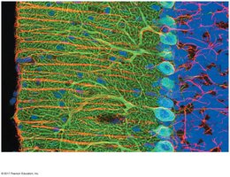
Example: The diversity of neuron shapes reflects their specialized functions in different regions of the nervous system.
Information Processing in Nervous Systems
Nervous systems process information in three main stages: sensory input, integration, and motor output.
Sensory Neurons: Detect external stimuli (e.g., light, touch, smell) and transmit information to the central nervous system (CNS).
Interneurons: Analyze and interpret sensory input, forming complex circuits within the CNS.
Motor Neurons: Transmit signals from the CNS to effector cells (muscles or glands), causing a response.
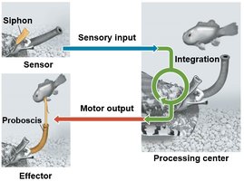
In animals, the central nervous system (CNS) includes the brain and spinal cord, while the peripheral nervous system (PNS) carries information to and from the CNS. Bundles of PNS neurons form nerves.
Ion Pumps, Ion Channels, and the Resting Potential
Establishing the Resting Potential
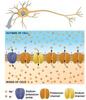
All cells maintain a voltage difference across their plasma membrane, known as the membrane potential. In neurons, the resting potential (typically -60 to -80 mV) is the membrane potential of a neuron not actively sending signals.
Sodium-Potassium Pump: Uses ATP to maintain high K+ inside and high Na+ outside the cell.
Ion Channels: Selectively allow ions to diffuse across the membrane, converting chemical potential to electrical potential.
At rest, many K+ channels are open, allowing K+ to diffuse out, creating a negative charge inside the cell.
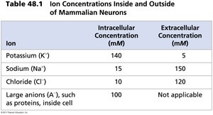
Ion | Intracellular Concentration (mM) | Extracellular Concentration (mM) |
|---|---|---|
Potassium (K+) | 140 | 5 |
Sodium (Na+) | 15 | 150 |
Chloride (Cl–) | 10 | 120 |
Large anions (A–) | 100 | Not applicable |
Example: The sodium-potassium pump and selective permeability of the membrane to K+ are essential for maintaining the resting potential.
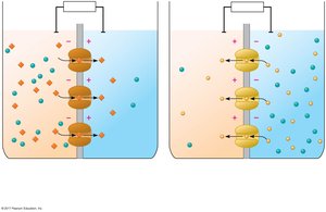
Modeling the Resting Potential
The resting potential can be modeled using an artificial membrane separating two chambers with different ion concentrations. K+ diffuses down its gradient, and the buildup of negative charge inside the cell balances the chemical and electrical gradients at equilibrium.
Action Potentials: The Signals of Neurons
Generation and Properties of Action Potentials
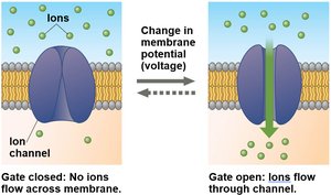
Neurons contain gated ion channels that open or close in response to stimuli, leading to changes in membrane potential. Voltage-gated ion channels open or close when the membrane potential reaches a certain threshold.
Graded Potentials: Small changes in membrane potential; magnitude varies with stimulus strength.
Action Potential: A rapid, all-or-none change in membrane voltage triggered when depolarization reaches threshold.

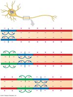
The action potential has a constant magnitude and transmits signals over long distances. It is generated at the axon hillock and travels toward the synaptic terminals.
Phases of the Action Potential
Resting State: Most voltage-gated Na+ and K+ channels are closed.
Depolarization: Stimulus opens some Na+ channels; if threshold is reached, more open, causing rapid depolarization.
Rising Phase: Na+ influx causes the inside to become positive.
Falling Phase: Na+ channels inactivate, K+ channels open, K+ exits, repolarizing the cell.
Undershoot: K+ channels remain open, causing hyperpolarization before returning to resting potential.
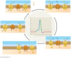
During the refractory period, a second action potential cannot be initiated due to inactivation of Na+ channels.
Conduction of Action Potentials
Action potentials propagate along the axon by depolarizing adjacent regions of the membrane. Inactivated Na+ channels behind the action potential prevent backward propagation.
Adaptations for Rapid Conduction
The speed of action potential conduction increases with axon diameter and the presence of a myelin sheath. In vertebrates, myelin sheaths are produced by oligodendrocytes (CNS) and Schwann cells (PNS). Action potentials jump between nodes of Ranvier in a process called saltatory conduction.
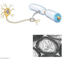
Synaptic Transmission
Types of Synapses
Neurons communicate with other cells at synapses, which can be electrical or chemical.
Electrical Synapses: Direct flow of current through gap junctions.
Chemical Synapses: Neurotransmitters carry information across the synaptic cleft.
At chemical synapses, the presynaptic neuron releases neurotransmitters stored in synaptic vesicles. The neurotransmitter diffuses across the synaptic cleft and binds to receptors on the postsynaptic cell.
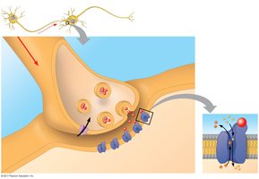
Postsynaptic Potentials and Summation
Neurotransmitter binding to ligand-gated ion channels generates postsynaptic potentials:
Excitatory Postsynaptic Potentials (EPSPs): Depolarizations that bring the membrane potential closer to threshold.
Inhibitory Postsynaptic Potentials (IPSPs): Hyperpolarizations that move the membrane potential farther from threshold.
Multiple EPSPs and IPSPs can combine through summation (temporal or spatial) to determine whether an action potential is generated in the postsynaptic neuron.
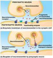
Termination of Neurotransmitter Signaling
Neurotransmitter signaling is terminated by:
Enzymatic breakdown of neurotransmitters in the synaptic cleft.
Reuptake of neurotransmitters by the presynaptic neuron.
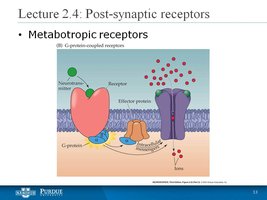
Modulated Signaling at Synapses
Some neurotransmitters bind to metabotropic receptors, activating signal transduction pathways involving second messengers. This can amplify the response and modulate the opening or closing of many ion channels.
Neurotransmitters
Major Classes of Neurotransmitters
Amino Acids: e.g., glutamate, GABA, glycine
Biogenic Amines: e.g., dopamine, serotonin, norepinephrine
Neuropeptides: e.g., substance P, endorphins
Acetylcholine: A key neurotransmitter at neuromuscular junctions
Gases: e.g., nitric oxide
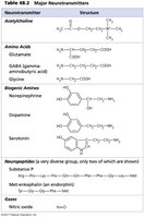
Example: Dopamine is involved in reward pathways and motor control; GABA is the main inhibitory neurotransmitter in the CNS.
Additional info: This guide expands on the provided slides with definitions, examples, and context for each concept, ensuring a comprehensive overview suitable for exam preparation in a college-level biology course.
