 Back
BackNeurons, Synapses, and Signaling: Structure and Function in the Nervous System
Study Guide - Smart Notes
 Tailored notes based on your materials, expanded with key definitions, examples, and context.
Neurons, Synapses, and Signaling
Key Concepts
Neuron organization and structure reflect function in information transfer.
Ion pumps and ion channels establish the resting membrane potential of a neuron.
Action potentials are the signals conducted by axons.
Neurons communicate with each other at synapses.
Neuron Structure and Function
Overview of Neurons
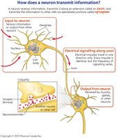
Neurons are specialized cells of the nervous system responsible for transmitting information throughout the body. Their structure is closely linked to their function, allowing for efficient communication via electrical and chemical signals.
Cell Body (Soma): Contains most of the neuron's organelles and is the metabolic center of the cell.
Dendrites: Highly branched extensions that receive signals from other neurons.
Axon: A long extension that transmits signals to other cells at synapses. The axon hillock is the cone-shaped base where action potentials are typically initiated.
Synaptic Terminal: The endpoint of an axon where neurotransmitters are released to communicate with other cells.
Types of Signals in Neurons
Neurons use two main types of signals:
Electrical signals: Used for long-distance communication along the axon.
Chemical signals: Used for short-distance communication at synapses via neurotransmitters.
Structural Diversity of Neurons
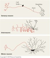
Neurons vary in structure depending on their function:
Sensory neurons: Transmit information from sensors that detect external or internal stimuli.
Interneurons: Integrate information within the brain or ganglia.
Motor neurons: Transmit signals to muscle or gland cells to elicit a response.
Glial Cells
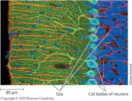
Most neurons are supported by glial cells, which provide nourishment, insulation, and structural support. In the mammalian brain, glia are essential for maintaining the environment around neurons and facilitating signal transmission.
Information Processing in the Nervous System
Stages of Information Processing
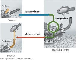
Nervous systems process information in three main stages:
Sensory Input: Sensors detect stimuli and transmit information via sensory neurons.
Integration: Interneurons in the brain or ganglia process and integrate sensory information.
Motor Output: Motor neurons carry signals from the integration centers to effectors (muscles or glands).
Organization of Nervous Systems
Central Nervous System (CNS): Includes the brain and nerve cord; site of information integration.
Peripheral Nervous System (PNS): Transmits information to and from the CNS; consists of bundled neurons called nerves.
Membrane Potentials and Ion Channels
Resting Membrane Potential
The resting membrane potential is the electrical potential difference across the neuron's plasma membrane when the cell is not transmitting a signal. It is established by the movement of ions, primarily sodium (Na+) and potassium (K+), through ion pumps and channels.
Ion Pumps: Actively transport ions against their concentration gradients (e.g., Na+/K+ pump).
Ion Channels: Allow passive movement of ions across the membrane; can be gated (open or close in response to stimuli).
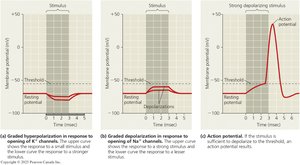
Hyperpolarization and Depolarization
Changes in membrane potential occur when gated ion channels open or close:
Depolarization: Membrane potential becomes less negative (e.g., Na+ enters the cell).
Hyperpolarization: Membrane potential becomes more negative (e.g., K+ leaves the cell).
Action Potentials
Graded Potentials vs. Action Potentials
Graded potentials are small changes in membrane potential whose magnitude varies with stimulus strength. If a depolarization reaches a threshold, it triggers an action potential—a rapid, all-or-none change in membrane voltage that travels along the axon.
Graded Potentials: Localized, variable in size, do not travel far.
Action Potentials: Large, uniform in size, propagate along the axon without loss of strength.
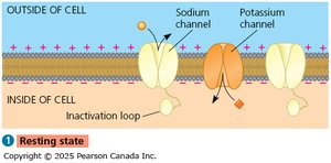
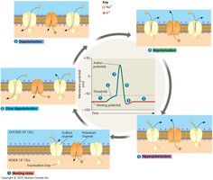
Generation of Action Potentials
The action potential is generated through a sequence of events involving voltage-gated Na+ and K+ channels:
At rest, most voltage-gated channels are closed.
Depolarization opens Na+ channels, allowing Na+ influx (rising phase).
Na+ channels inactivate; K+ channels open, allowing K+ efflux (falling phase).
During the undershoot, K+ permeability is high, then returns to resting levels.
The refractory period follows, during which a second action potential cannot be initiated due to Na+ channel inactivation.
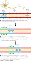
Conduction of Action Potentials
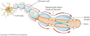
Propagation Along the Axon
Action potentials are typically initiated at the axon hillock and propagate in one direction toward the synaptic terminals. This unidirectional flow is ensured by the inactivation of Na+ channels behind the depolarization zone.
Adaptations for Speed
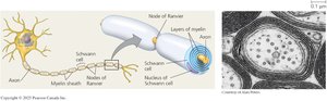
The speed of action potential conduction is influenced by axon diameter and the presence of a myelin sheath:
Larger diameter: Faster conduction due to lower resistance.
Myelin sheath: Insulates axons, allowing for saltatory conduction, where action potentials jump between nodes of Ranvier (gaps in the myelin sheath).
Glial cells: Oligodendrocytes (CNS) and Schwann cells (PNS) produce myelin.
Synaptic Transmission
Types of Synapses
Neurons communicate with other cells at specialized junctions called synapses. There are two main types:
Electrical synapses: Direct flow of electrical current via gap junctions.
Chemical synapses: Use neurotransmitters to transmit signals across a synaptic cleft; most common type in the nervous system.
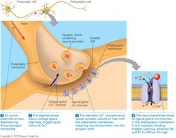
Chemical Synapse Mechanism
At a chemical synapse, the arrival of an action potential at the synaptic terminal triggers the release of neurotransmitters, which cross the synaptic cleft and bind to receptors on the postsynaptic cell, initiating a response.
Summary Table: Key Features of Neurons and Synaptic Signaling
Feature | Description | Example/Function |
|---|---|---|
Neuron Structure | Cell body, dendrites, axon, synaptic terminal | Signal reception, integration, transmission |
Glial Cells | Support, nourish, insulate neurons | Oligodendrocytes, Schwann cells |
Resting Potential | Electrical potential across membrane at rest | ~ -70 mV in typical neuron |
Action Potential | All-or-none electrical signal | Long-distance communication |
Synapse | Junction between neurons or neuron and effector | Chemical (neurotransmitter) or electrical (gap junction) |
Key Equations
Nernst Equation (for equilibrium potential of an ion): $E_{ion} = \frac{RT}{zF} \ln \left( \frac{[ion]_{outside}}{[ion]_{inside}} \right)$
Resting Membrane Potential (Goldman-Hodgkin-Katz equation): $V_m = \frac{RT}{F} \ln \left( \frac{P_{K^+}[K^+]_{out} + P_{Na^+}[Na^+]_{out} + P_{Cl^-}[Cl^-]_{in}}{P_{K^+}[K^+]_{in} + P_{Na^+}[Na^+]_{in} + P_{Cl^-}[Cl^-]_{out}} \right)$
Additional info: The above equations are fundamental for understanding how ion gradients and membrane permeability contribute to the electrical properties of neurons.
