 Back
BackNeurons, Synapses, and Signaling: Structure and Function in Information Transfer
Study Guide - Smart Notes
 Tailored notes based on your materials, expanded with key definitions, examples, and context.
Neurons, Synapses, and Signaling
Introduction to Neurons and Nervous System Function
The nervous system is responsible for the rapid transmission and processing of information in animals. Neurons are the fundamental units of the nervous system, specialized for the conduction of electrical signals and communication with other cells.
Neurons are excitable cells that transmit information via electrical and chemical signals.
Glia (or glial cells) support, nourish, and insulate neurons.
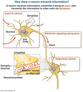
Information is transmitted from a presynaptic cell (usually a neuron) to a postsynaptic cell (neuron, muscle, or gland cell) at specialized junctions called synapses.
Neuron Structure and Types
Neurons have specialized structures that reflect their function in information transfer:
Dendrites: Receive incoming signals from other neurons.
Cell body (soma): Contains the nucleus and organelles; integrates incoming signals.
Axon: Conducts electrical impulses away from the cell body toward synaptic terminals.
Synaptic terminals: Release neurotransmitters to communicate with postsynaptic cells.
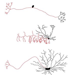
Neurons can be classified by structure and function:
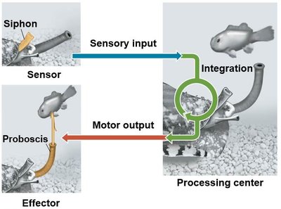
Sensory neurons: Transmit information about external stimuli (e.g., light, touch, smell).
Interneurons: Integrate and interpret information within the central nervous system (CNS).
Motor neurons: Transmit signals to muscle cells, causing contraction.
Organization of the Nervous System
Many animals have a complex nervous system with two main divisions:
Central Nervous System (CNS): Site of integration; includes the brain and nerve cord.
Peripheral Nervous System (PNS): Carries information into and out of the CNS; neurons in the PNS are bundled into nerves.
Ion Basis of Neuronal Signaling
Membrane Potential and Resting Potential
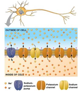
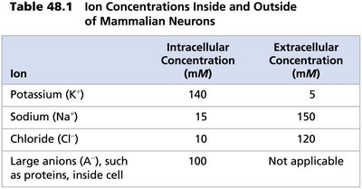
Every cell has a voltage difference across its plasma membrane, known as the membrane potential. In neurons, the resting potential is the membrane potential of a neuron not sending signals, typically around -70 mV.
Ion gradients are established by sodium-potassium pumps, which use ATP to maintain high K+ inside and high Na+ outside the cell.
These gradients represent chemical potential energy.
Ion | Intracellular Concentration (mM) | Extracellular Concentration (mM) |
|---|---|---|
Potassium (K+) | 140 | 5 |
Sodium (Na+) | 15 | 150 |
Chloride (Cl–) | 10 | 120 |
Large anions (A–) | 100 | Not applicable |
Establishment of Resting Potential
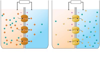
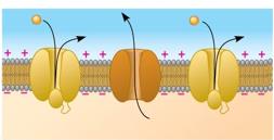
The opening of ion channels in the plasma membrane converts chemical potential to electrical potential. At rest, many K+ channels are open, allowing K+ to diffuse out, resulting in a net negative charge inside the neuron.
Gated Ion Channels and Changes in Membrane Potential
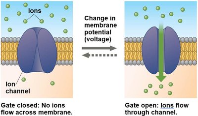
Neurons contain gated ion channels that open or close in response to stimuli, altering the membrane potential. Voltage-gated ion channels respond to changes in membrane voltage.
Action Potentials
Generation and Properties of Action Potentials
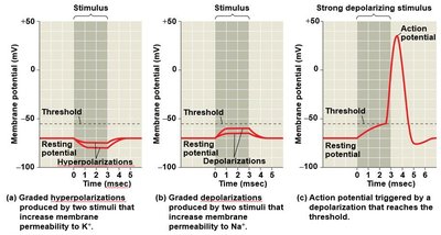
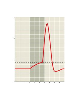
An action potential is a rapid, all-or-none change in membrane potential that transmits signals along the axon. It is triggered when depolarization reaches a threshold value.
Graded potentials: Small changes in polarization; magnitude varies with stimulus strength.
Action potentials: Large, constant-magnitude changes; all-or-none response.
Phases of the Action Potential
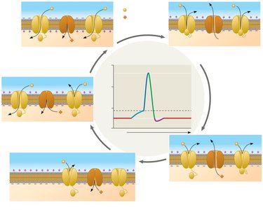
Resting state: Most voltage-gated Na+ and K+ channels are closed.
Depolarization: Na+ channels open, Na+ enters the cell.
Rising phase: Rapid influx of Na+ causes membrane potential to become positive.
Falling phase: Na+ channels inactivate, K+ channels open, K+ exits the cell.
Undershoot: Membrane potential temporarily becomes more negative than resting potential.
Refractory Period
After an action potential, a refractory period occurs during which a second action potential cannot be initiated. This is due to temporary inactivation of Na+ channels.
Conduction of Action Potentials
Action potentials are generated at the axon hillock and travel in one direction toward the synaptic terminals. Inactivated Na+ channels behind the depolarization zone prevent backward propagation.
Adaptations for Rapid Conduction
Action potential speed increases with axon diameter (invertebrates have thick axons).
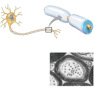
In vertebrates, myelin sheaths (produced by oligodendrocytes in the CNS and Schwann cells in the PNS) insulate axons, increasing conduction speed.
Action potentials jump between nodes of Ranvier in a process called saltatory conduction.
Synaptic Transmission
Types of Synapses
Electrical synapses: Electrical current flows directly between cells via gap junctions.
Chemical synapses: Neurotransmitters carry information across the synaptic cleft.
Most synapses in the nervous system are chemical synapses.
Chemical Synaptic Transmission
The presynaptic neuron synthesizes and packages neurotransmitters in synaptic vesicles.
An action potential triggers neurotransmitter release into the synaptic cleft.
Neurotransmitters bind to receptors on the postsynaptic cell, generating a postsynaptic potential.
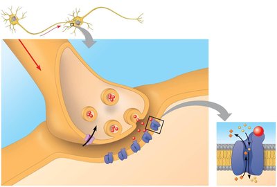
Postsynaptic Potentials and Summation
Excitatory postsynaptic potentials (EPSPs): Depolarizations that bring the membrane potential toward threshold.
Inhibitory postsynaptic potentials (IPSPs): Hyperpolarizations that move the membrane potential farther from threshold.
Summation: Multiple EPSPs and IPSPs can combine (spatially or temporally) to determine whether an action potential is generated.
Termination of Neurotransmitter Signaling
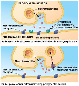
Neurotransmitter signaling is terminated by enzymatic breakdown or reuptake into the presynaptic cell.
Modulation of Synaptic Signaling
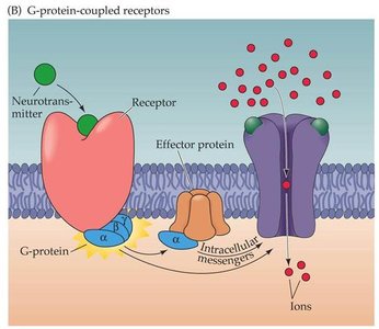
Some neurotransmitters bind to metabotropic receptors, activating signal transduction pathways involving second messengers.
This can amplify the response, opening or closing many ion channels.
Major Neurotransmitters
Acetylcholine: Involved in muscle stimulation, memory, and learning.
Amino acids: Glutamate (excitatory), GABA (inhibitory), glycine.
Biogenic amines: Norepinephrine, dopamine, serotonin (involved in mood, attention, and learning).
Neuropeptides: Substance P, endorphins (modulate pain perception).
Gases: Nitric oxide (NO) acts as a local regulator.
Neurotransmitter | Structure |
|---|---|
Acetylcholine | Structure shown in original table |
Glutamate | Structure shown in original table |
GABA | Structure shown in original table |
Glycine | Structure shown in original table |
Norepinephrine | Structure shown in original table |
Dopamine | Structure shown in original table |
Serotonin | Structure shown in original table |
Substance P | Structure shown in original table |
Met-enkephalin | Structure shown in original table |
Nitric oxide | NO |
Example: Acetylcholine is released at neuromuscular junctions, causing muscle contraction. GABA is the main inhibitory neurotransmitter in the brain.
Additional info: The diversity of neurotransmitters and their receptors allows for complex modulation and integration of nervous system signals.
