 Back
BackNeurons, Synapses, and Signaling: Structure and Function of the Nervous System
Study Guide - Smart Notes
 Tailored notes based on your materials, expanded with key definitions, examples, and context.
Neurons, Synapses, and Signaling
Overview of Neuronal Function
Neurons are the fundamental units of the nervous system, specialized for the reception, transmission, and integration of information. They use electrical impulses and chemical signals to communicate over both short and long distances, enabling complex processes such as sensation, movement, learning, and memory.
Neurons transmit information using electrical impulses (action potentials) and chemical messengers (neurotransmitters).
Neuronal communication is essential for physiological responses and behavioral outputs.
Neurons are organized into networks, with complex animals possessing centralized processing centers such as brains or ganglia.
Stages of Information Processing
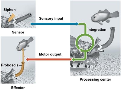
Neural information processing involves four main stages: sensory input, integration, motor output, and learning/memory.
Sensory Input: Detection of external or internal stimuli (e.g., light, blood pressure).
Integration: Processing and interpretation of sensory information, often within the central nervous system (CNS).
Motor Output: Transmission of signals to effectors (muscles or glands) to elicit a response.
Learning & Memory: Modification of responses based on experience.
Structure of Neurons
Basic Anatomy of a Neuron
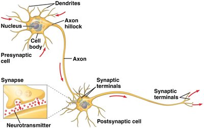
Neurons have specialized structures for receiving, integrating, and transmitting information.
Dendrites: Branched extensions that receive signals from other neurons.
Cell Body (Soma): Contains the nucleus and organelles; integrates incoming signals.
Axon Hillock: Region where action potentials are initiated.
Axon: Long projection that conducts action potentials away from the cell body.
Synaptic Terminals: Branched endings that transmit signals to other cells at synapses.
Synapse: Junction where information is transferred to another cell, often via neurotransmitters.
Types of Neurons
Neurons are classified based on their function:
Sensory Neurons: Transmit information about external and internal stimuli to the CNS.
Interneurons: Integrate information within the CNS; form complex circuits.
Motor Neurons: Convey signals from the CNS to effectors (muscles or glands).
Organization of the Nervous System
The nervous system is divided into the central nervous system (CNS) and peripheral nervous system (PNS):
CNS: Brain and spinal cord; site of integration.
PNS: Nerves and ganglia outside the CNS; transmits sensory and motor information.
Nerves: Bundles of axons in the PNS.
Glial Cells
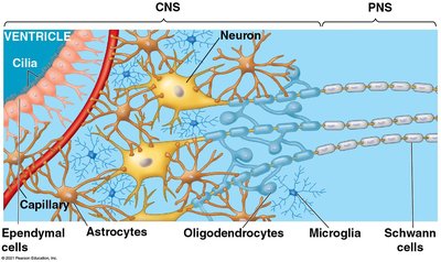
Glial cells support and protect neurons. Types include:
Astrocytes: Facilitate information transfer, form the blood-brain barrier, and nourish neurons.
Oligodendrocytes (CNS) and Schwann Cells (PNS): Myelinate axons, increasing conduction speed.
Microglia: Immune defense in the CNS.
Ependymal Cells: Line brain ventricles and help circulate cerebrospinal fluid.
Membrane Potential and Resting Potential
Establishment of Resting Potential
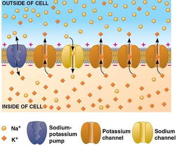
The resting potential is the voltage difference across the plasma membrane of a neuron at rest, typically between -60 and -80 mV. It is established by the unequal distribution of ions and selective permeability of the membrane.
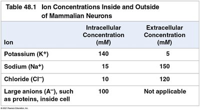
Key Ions: Potassium (K+), Sodium (Na+), Chloride (Cl-), and large anions (A-).
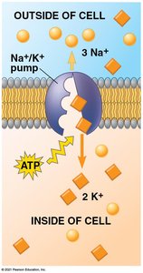
Ion Gradients: Maintained by the sodium-potassium pump (3 Na+ out, 2 K+ in per ATP hydrolyzed).
K+ Leak Channels: Allow K+ to move out, making the inside more negative.
Na+ Leak Channels: Few in number; allow some Na+ influx, making the resting potential less negative than the K+ equilibrium potential.
Ion | Intracellular Concentration (mM) | Extracellular Concentration (mM) |
|---|---|---|
Potassium (K+) | 140 | 5 |
Sodium (Na+) | 15 | 150 |
Chloride (Cl-) | 10 | 120 |
Large anions (A-) | 100 | Not applicable |
The Nernst Equation and Equilibrium Potential
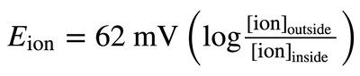
The equilibrium potential for an ion is the membrane voltage at which the net flow of that ion across the membrane is zero. It can be calculated using the Nernst equation:

For K+, the equilibrium potential is about -90 mV; for Na+, it is about +62 mV. The actual resting potential is closer to K+'s equilibrium potential due to higher permeability to K+.
Changes in Membrane Potential: Graded and Action Potentials
Gated Ion Channels and Graded Potentials
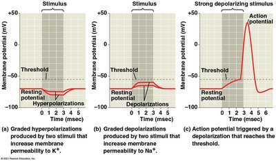
Neurons respond to stimuli by opening gated ion channels, causing changes in membrane potential:
Hyperpolarization: Opening K+ channels increases negativity (toward -90 mV).
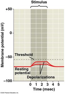
Depolarization: Opening Na+ channels decreases negativity (toward +62 mV).
Graded Potentials: Small, local changes in membrane potential that decay with distance.
Action Potentials
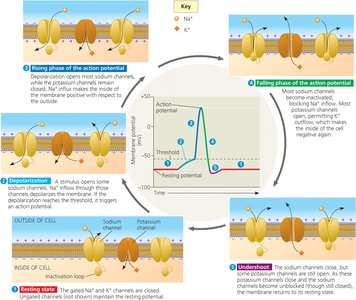
An action potential is a rapid, all-or-none electrical signal that travels along the axon. It is triggered when depolarization reaches a threshold (about -55 mV in mammals).
Phases: Depolarization (Na+ influx), repolarization (K+ efflux), and hyperpolarization.
Refractory Period: Brief inactivation of Na+ channels prevents backward propagation.
Conduction: Action potentials are regenerated along the axon, ensuring signal fidelity.
Saltatory vs. Continuous Conduction
Myelinated axons (in vertebrates) conduct action potentials more rapidly via saltatory conduction, where the impulse jumps between nodes of Ranvier. Unmyelinated axons (in invertebrates) conduct signals continuously and more slowly.
Saltatory Conduction: Myelin sheaths insulate axons; action potentials jump between nodes.
Continuous Conduction: Action potentials propagate along the entire axon membrane.
Synaptic Transmission
Chemical and Electrical Synapses
Neurons communicate at synapses, which can be chemical or electrical:
Chemical Synapses: Neurotransmitters are released from the presynaptic neuron and bind to receptors on the postsynaptic cell, generating postsynaptic potentials.
Electrical Synapses: Direct flow of ions through gap junctions; allow rapid, synchronized activity.
Postsynaptic Potentials
Neurotransmitter binding can cause excitatory or inhibitory postsynaptic potentials:
Excitatory Postsynaptic Potential (EPSP): Depolarizes the postsynaptic membrane, increasing the likelihood of an action potential.
Inhibitory Postsynaptic Potential (IPSP): Hyperpolarizes the membrane, decreasing the likelihood of an action potential.
Summation and Signal Integration
Multiple postsynaptic potentials can summate:
Temporal Summation: Rapid EPSPs at the same synapse add together.
Spatial Summation: EPSPs from different synapses combine.
IPSPs: Can negate EPSPs, preventing action potential generation.
Major Neurotransmitters and Signal Termination
Acetylcholine: Muscle stimulation, memory, learning; effects depend on receptor type.
Glutamate: Main excitatory neurotransmitter in the CNS; involved in memory formation.
Dopamine and Serotonin: Modulate mood, attention, and learning.
Nitric Oxide: Relaxes smooth muscle.
Signal Termination: Neurotransmitters are removed by enzymatic breakdown, reuptake, or diffusion.
Summary
Neurons use electrical and chemical signals to process and transmit information.
Resting potential is established by ion gradients and selective permeability.
Action potentials are all-or-none signals that propagate along axons.
Synaptic transmission enables communication between neurons and target cells.
