 Back
BackNeurons, Synapses, and Signaling: Structure and Function of the Nervous System
Study Guide - Smart Notes
 Tailored notes based on your materials, expanded with key definitions, examples, and context.
Neurons, Synapses, and Signaling
Neuron Organization and Structure Reflect Function in Information Transfer
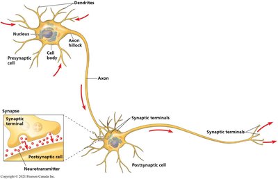
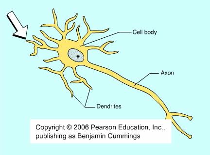
Neurons are specialized cells of the nervous system responsible for transmitting information throughout the body. Their structure is closely related to their function, with distinct regions for receiving, integrating, and transmitting signals.
Cell Body (Soma): Contains the nucleus and most organelles.
Dendrites: Highly branched extensions that receive signals from other neurons.
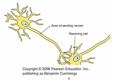
Axon: A long extension that transmits signals to other cells, often ending in multiple synaptic terminals.
Axon Hillock: The cone-shaped base of the axon where action potentials are typically initiated.
Synapse: The junction between the axon terminal of one neuron and the target cell (another neuron, muscle, or gland cell).
Neurotransmitters: Chemical messengers released from synaptic terminals to transmit signals across the synapse.
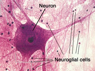
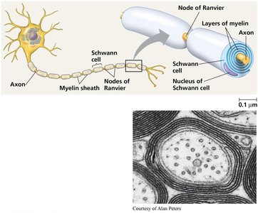
Glial Cells: Non-neuronal cells that nourish, insulate, and support neurons. Types include astrocytes, oligodendrocytes, and Schwann cells.
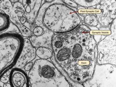
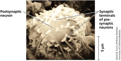
Synaptic Structure: Electron micrographs reveal synaptic vesicles in the presynaptic terminal, the synaptic cleft, and the postsynaptic cell membrane.
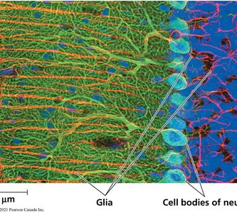
Glia in Nervous Tissue: Glial cells form networks that support and interact with neurons.
Introduction to Information Processing
Nervous systems process information in three main stages:
Sensory Input: Sensors detect external and internal stimuli and transmit information via sensory neurons.
Integration: Interneurons in the brain or ganglia process and integrate sensory input.
Motor Output: Motor neurons carry signals from the CNS to effectors (muscles or glands) to elicit a response.
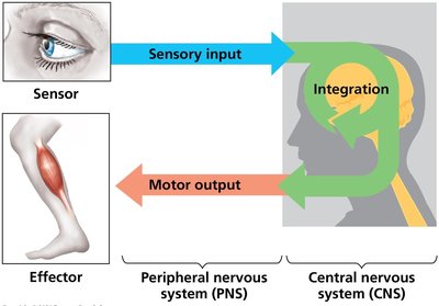
The nervous system is divided into the central nervous system (CNS)—brain and spinal cord—and the peripheral nervous system (PNS), which carries information to and from the CNS. Bundles of PNS neurons form nerves.
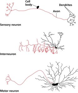
Neurons are classified by function and structure:
Sensory Neurons: Transmit sensory information to the CNS.
Interneurons: Integrate information within the CNS.
Motor Neurons: Transmit signals from the CNS to effectors.
Ion Pumps and Ion Channels Establish the Resting Membrane Potential of a Neuron
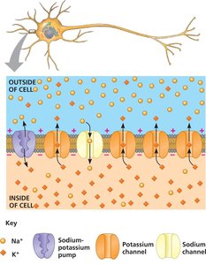
All cells maintain a voltage difference across their plasma membrane, known as the membrane potential. In neurons, the resting potential is typically around -70 mV, with the inside of the cell more negative than the outside.
Sodium-Potassium Pump: Uses ATP to maintain high K+ inside and high Na+ outside the cell.
Ion Channels: Selectively allow ions to diffuse across the membrane, converting chemical potential to electrical potential.
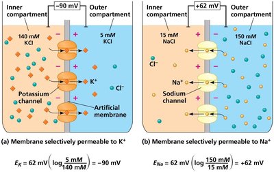
The Nernst equation calculates the equilibrium potential for a particular ion:
$E_{ion} = 62\ \mathrm{mV} \times \log \left( \frac{[\text{ion}]_{\text{outside}}}{[\text{ion}]_{\text{inside}}} \right)$
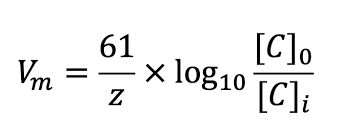
Generalized Nernst equation:
$V_m = \frac{61}{z} \times \log_{10} \frac{[C]_0}{[C]_i}$
Action Potentials: Signals Conducted by Axons
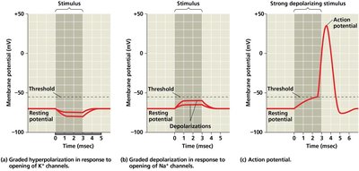
Neurons communicate via changes in membrane potential. Gated ion channels open or close in response to stimuli, leading to graded potentials or action potentials.
Depolarization: Membrane potential becomes less negative (e.g., Na+ influx).
Hyperpolarization: Membrane potential becomes more negative (e.g., K+ efflux).
Graded Potentials: Small changes in membrane potential; magnitude varies with stimulus strength.
Action Potentials: Large, all-or-none changes in membrane potential that propagate along the axon.
Generation of Action Potentials
Action potentials occur in several stages:
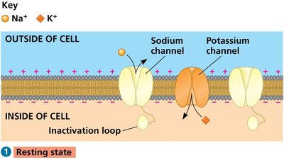
Resting State: Most voltage-gated Na+ and K+ channels are closed.
Depolarization: Voltage-gated Na+ channels open, Na+ enters the cell.
Rising Phase: Membrane potential rapidly increases as more Na+ channels open.
Falling Phase: Na+ channels inactivate, K+ channels open, K+ exits the cell.
Undershoot: K+ permeability is temporarily higher than at rest, then returns to baseline.
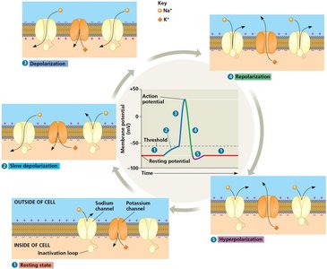
During the refractory period, a second action potential cannot be initiated due to inactivation of Na+ channels.
Conduction of Action Potentials
Action potentials are typically initiated at the axon hillock and propagate unidirectionally toward synaptic terminals. Inactivated Na+ channels prevent backward propagation.
Evolutionary Adaptation of Axon Structure
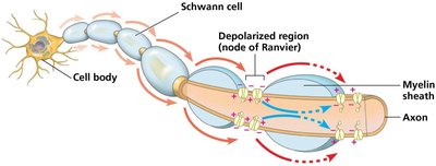
The speed of action potential conduction increases with axon diameter and the presence of a myelin sheath. Myelin is produced by oligodendrocytes (CNS) and Schwann cells (PNS).

In myelinated axons, action potentials jump between nodes of Ranvier in a process called saltatory conduction.
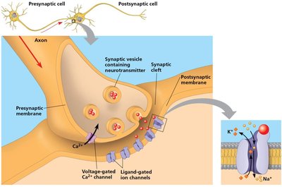
Neurons Communicate with Each Other at Synapses
Neurons communicate at synapses, which can be electrical (via gap junctions) or chemical (via neurotransmitters). Most synapses in the nervous system are chemical.
At chemical synapses:
The presynaptic neuron releases neurotransmitter from synaptic vesicles into the synaptic cleft.
The neurotransmitter binds to receptors on the postsynaptic cell, causing ion channels to open and generating a postsynaptic potential.
Generation and Summation of Postsynaptic Potentials
Postsynaptic potentials can be:
Excitatory (EPSPs): Depolarize the postsynaptic membrane, bringing it closer to threshold.
Inhibitory (IPSPs): Hyperpolarize the membrane, moving it further from threshold.
Summation of EPSPs and IPSPs determines whether the postsynaptic neuron will fire an action potential:
Temporal Summation: Multiple EPSPs from a single synapse in rapid succession add together.
Spatial Summation: EPSPs from multiple synapses on the same neuron combine.
IPSPs can counteract EPSPs, and the net effect at the axon hillock determines action potential generation.
Summary Table: Ion Concentrations Inside and Outside of Mammalian Neurons
Ion | Intracellular Concentration (mM) | Extracellular Concentration (mM) |
|---|---|---|
Potassium (K+) | 140 | 5 |
Sodium (Na+) | 15 | 150 |
Chloride (Cl−) | 10 | 120 |
Large anions (A−) | 100 | Not applicable |
Additional info: This table highlights the ionic gradients maintained by neurons, which are essential for generating membrane potentials and action potentials.
