 Back
Neurons, Synapses, and Signaling: Structure and Function in the Nervous System
Study Guide - Smart Notes
 Tailored notes based on your materials, expanded with key definitions, examples, and context.
Neurons, Synapses, and Signaling
Lines of Communication in the Nervous System
The nervous system relies on specialized cells called neurons to transfer information throughout the body. Neurons communicate using both electrical and chemical signals, allowing for rapid and precise control of bodily functions.
Electrical signals travel over long distances within neurons.
Chemical signals operate over short distances between neurons or between neurons and other cells.
Neurons are organized into clusters for processing information:
Ganglia: Simple clusters for basic processing.
Brain: Complex clusters for advanced processing.
Neuron Structure
Neurons have specialized structures that enable them to receive, process, and transmit information efficiently.
Cell body: Contains most of the neuron's organelles and is the metabolic center.
Dendrites: Highly branched extensions that receive incoming signals from other neurons.
Axon: A long extension that transmits signals to other cells; axons often branch at their ends to connect with multiple target cells.
Synapse: The junction between the axon terminal of one neuron and the dendrite or cell body of another; the site of signal transmission.
Neurotransmitters: Chemical messengers that cross the synaptic gap to transmit information from one neuron to another.
Neuron Support: Glial Cells
Neurons require support from glial cells, which perform various essential functions in the nervous system.
Glial cells: Provide energy, insulation, and immune responses; outnumber neurons in the mammalian brain by 10–50:1.
Astrocytes: Maintain nutrient reserves, form scar tissue, and can act as stem cells.
Oligodendrocytes: Form the myelin sheath in the central nervous system (CNS).
Schwann cells: Form the myelin sheath in the peripheral nervous system (PNS).
Microglial cells: Act as immune cells in the CNS by phagocytosing debris and pathogens.
Membrane Potential and Action Potentials
Ion Pumps and Channels
The membrane potential is the electrical charge difference across the plasma membrane, established by ion pumps and channels. This potential is essential for the generation and propagation of electrical signals in neurons.
Resting potential: The steady-state difference in charge across the membrane, primarily maintained by potassium (K+) and sodium (Na+) ions.
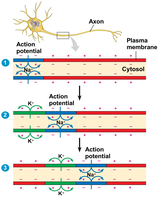
Action potential: A rapid, temporary change in membrane potential in response to a stimulus, allowing the signal to travel along the neuron.
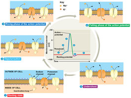
Transport of Na+ and K+ in Neurons
Neurons use specialized proteins to move sodium and potassium ions across their membranes, which is critical for generating action potentials.
Sodium-potassium pump: Actively transports 3 Na+ ions out and 2 K+ ions into the cell, maintaining the resting potential.
Voltage-gated channels: Open or close in response to changes in membrane potential, allowing for rapid ion movement during action potentials.
Phases of the Action Potential
The action potential consists of several distinct phases, each involving specific changes in ion permeability and membrane potential.
Depolarization: Na+ channels open, allowing Na+ to enter the cell, making the inside more positive.
Rising phase: Rapid influx of Na+ causes a sharp increase in membrane potential.
Falling phase: Na+ channels inactivate, K+ channels open, and K+ exits the cell, restoring negativity.
Undershoot: Membrane potential temporarily becomes more negative than resting potential due to continued K+ efflux.
Refractory period: Brief period (1–2 ms) during which the neuron cannot fire another action potential, ensuring unidirectional signal transmission.
Synapses and Neurotransmission
Types of Synapses
Neurons communicate with other cells at specialized junctions called synapses. There are two main types:
Electrical synapse: Direct cytoplasmic connections (gap junctions) allow ions to flow directly between cells, enabling rapid signal transmission.
Chemical synapse: A physical gap (synaptic cleft) separates cells; neurotransmitters are released from the presynaptic neuron and bind to receptors on the postsynaptic cell.
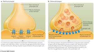
Synaptic Transmission and Neurotransmitters
At chemical synapses, neurotransmitters mediate communication between neurons. The response depends on both the neurotransmitter released and the receptors present on the postsynaptic cell.
Neurotransmitter release: Triggered by the arrival of an action potential, causing Ca2+ influx and vesicle fusion with the presynaptic membrane.
Receptor diversity: A single neurotransmitter can have multiple receptor types, leading to varied effects.
Acetylcholine: Involved in arousal, attention, memory, motivation, and muscle activation.
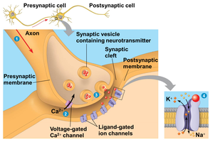
Information Processing in the Nervous System
Main Steps of Information Processing
The nervous system processes information in three main stages:
Sensory input: Detection of stimuli by sensory receptors.
Integration: Processing and interpretation of sensory input by the central nervous system (CNS).
Motor output: Transmission of signals to effector cells (muscles or glands) via the peripheral nervous system (PNS).
Types of Neurons
Neurons are classified based on their function in information processing:
Afferent neurons (sensory neurons): Receive information from sensory receptors and transmit it to the CNS.
Interneurons: Integrate sensory input and formulate responses within the CNS.
Efferent neurons (motor neurons): Carry out the response by transmitting signals from the CNS to effectors.
